Unsupported Browser
The American College of Surgeons website is not compatible with Internet Explorer 11, IE 11. For the best experience please update your browser.
Menu
Become a member and receive career-enhancing benefits

Our top priority is providing value to members. Your Member Services team is here to ensure you maximize your ACS member benefits, participate in College activities, and engage with your ACS colleagues. It's all here.

Become a Member
Become a member and receive career-enhancing benefits

Our top priority is providing value to members. Your Member Services team is here to ensure you maximize your ACS member benefits, participate in College activities, and engage with your ACS colleagues. It's all here.

Become a Member
ACS
Case Reviews in Surgery

Laparoscopic Left Hepatectomy and Common Bile Duct Exploration for Recurrent Pyogenic Cholangiohepatitis: A Case Demonstrating the Use of Cognitive Task Analysis to Create a Step Wise Approach to Laparoscopic Liver Surgery

Laparoscopic Left Hepatectomy and Common Bile Duct Exploration for Recurrent Pyogenic Cholangiohepatitis: A Case Demonstrating the Use of Cognitive Task Analysis to Create a Step Wise Approach to Laparoscopic Liver Surgery

March 1, 2021

Abstract

Background

Recurrent episodes of cholangiohepatitis due to pyogenic bile duct infection have an associated high morbidity and mortality.

Summary

A 42-year-old male of Korean descent presented with recurrent pyogenic cholangitis (RPC) secondary to a left intrahepatic bile duct stricture. The patient also suffered from recurrent intrahepatic cholelithiasis. The patient was initially managed with six endoscopic retrograde cholangio-pancreatography (ERCP) procedures involving stent placement and exchanges for stricture management over six months without improvement. RPC is a challenging surgical disease to manage and evidence supports hepatectomy for definitive management. The patient underwent a laparoscopic left hepatectomy and left common bile duct exploration—this operation relies on safe hepatic transection. A step-wise operative technique of taking the artery, portal vein, and then bile duct to the resected specimen using the caudal approach is outlined. In addition, ultrasound mapping to navigate the liver and using the middle vein as the endpoint for parenchymal transection to avoid getting lost in the liver is described. This case description highlights cautionary points, pitfalls, and tips for success in this technically challenging operation.

Conclusion

Definitive management of recurrent pyogenic cholangiohepatitis via a laparoscopic hepatectomy with common bile duct exploration is a safe and effective approach. Cognitive task analysis can be used to break down a complex operation into modular tasks to educate surgeons how to incorporate each task in their practice or improve their current approach to laparoscopic liver surgery.

Key Words

Laparoscopic liver surgery, recurrent pyogenic cholangiohepatitis, cognitive task analysis, common bile duct exploration


Case Description

This case report demonstrates the use of cognitive task analysis, which has been shown to improve procedural learning, to break down a complex operation into modular tasks.1-4 The intent of this method is to educate surgeons how to incorporate each task in their practice or improve their current approach to laparoscopic liver surgery. Experts may use this outline to improve teaching skills by breaking down an automated psychomotor task into discrete steps.

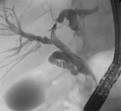
A 42-year-old Korean male presented with a one-year history of chronic, left-sided RPC resulting in a left hepatic duct stricture which only worsened the patient’s biliary disease. The patient’s treatment course included six prior ERCPs for stent placement and exchanges without improvement. At six-month follow-up, ERCP continued to show disease in the left hepatic duct (Figure 1). Unfortunately, this is a common disease trajectory for RPC and the patient was counseled to undergo hepatic resection.

Figure 1. ERCP demonstrating a persistent stricture in the left hepatic duct with post-stricture biliary ductal dilatation.

Step 1: Liver Mobilization

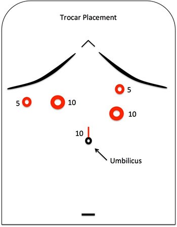
The operation begins with proper trocar placement (Figure 2). The right mid-clavicular 10 mm port site is placed higher than usual because the cavitron ultrasonic surgical aspirator (CUSA) instrument, used for parenchymal dissection, is short and needs to be used through this site. After abdominal exploration, liver mobilization is achieved by mobilization of the suprahepatic vena cava, left coronary, and triangular ligaments. For this case, we planned to leave the middle hepatic vein (MHV), thus the bifurcation between the MHV and left hepatic vein (LHV) was identified with intraoperative ultrasound and marked on the liver. This point serves as the endpoint for transection.

Figure 2. Proper trocar placement when planning for a laparoscopic liver resection. Notice that the 10mm port in the right midclavicular position is placed higher than usual to accommodate the short CUSA instrument.

Step 2: Remove gallbladder adhesions

Dense adhesions to the gallbladder are then taken down. These adhesions usually occur as a result of repeated episodes of cholecystitis or cholangitis. Bleeding is common due to active inflammation and fusion of tissues. It is important not to leave omentum on the gallbladder. Some tissue surface bleeding is often encountered, due to the inflammatory response, and bipolar cautery is ideal to control such bleeding.

Step 3: Isolate the hepatoduodenal ligament

The next step is isolation of the hepatoduodenal ligament (HDL). This allows the surgeon to perform a Pringle maneuver during parenchymal transection. Often, the duodenum is adhered to the anterior surface of the HDL. Because of this, the surgeon should start by entering the lesser sac and identify the spigelian lobe. Then, dissection of the right lateral aspect of the HDL down to the inferior vena cava is carried out. Now the anterior duodenum can be safely dissected off the HDL, as landmarks are clearly visible, and then the foramen of Winslow can be recreated. At this time a soft umbilical tape can be placed and used for Pringle maneuver when needed.

Step 4: Hepatoduodenal ligament dissection

The fourth step is to start the dissection of the hepatoduodenal ligament. This starts with identification of the hepatic arterial system. The surgeon must identify the bifurcation of the left and right hepatic arteries in order to be confident that the correct artery is transected to the specimen. This requires delicate dissection in order to isolate the bifurcation (Figure 3).

Figure 3. Demonstration of the hepatic arterial bifurcation during hepatoduodenal ligament dissection.

Proper dissection of the hepatic arterial system and taking of the left hepatic artery with a vascular stapler or clips is essential to facilitating exposure, isolation, and transection of the left portal vein.

Isolation of the left portal vein is then approached and this may require taking down a caudate branch. The left portal vein is isolated and clamped with a bulldog. Lack of flow to the left hepatic lobe is confirmed and presence of flow to the right liver is confirmed using intraoperative ultrasound. The left portal vein can then be taken with a stapler.

For biliary disease, it is not recommended to isolate or transect the bile duct at this time because when performing liver resection for biliary disease, extra-fascial dissection is not recommended. Inflamed tissue can be a safety hazard due to potential adherence of structures and possible misidentification of right-sided structures, specifically the right posterior section bile duct can insert on the left transverse bile duct. This step is delayed until later (see below).

Step 5: Preparing for hepatic transection

Preparation for hepatic transection is essential for safe liver surgery. Two aspects are critical: The first is ultrasound mapping and marking on the surface of the liver. This should be done from the bifurcation of the MHV-LHV all the way down to the hilum of the liver. In this case, marking to the left side of the MHV was performed because of the intention to leave the MHV in situ. The second step is to create a 1.5 cm deep crevasse into the liver, using a harmonic dissector, along this pre-marked line (Figure 4). This is essential during the caudal approach because during transection, vision is limited (no anterior/global view), and the crevasse will serve as a transection guide (Figure 5).

Figure 4. Initiation of the crevasse along the pre-marked transection line, planned using ultrasound from the suprahepatic IVC down to the hilum of the liver.

Figure 5. Demonstration of the 1.5cm delineated crevasse serving as the parenchymal transection guide during hepatic transection.

Step 6: Hepatic transection using the caudal approach

The next step is hepatic transection using the caudal approach.5-7 The Pringle should be applied and the CUSA is used to dissect the parenchyma. As structures are identified, they are transected using a combination of clips, energy devices, and staplers (rarely). The delineated crevasse serves as the line of transection and helps the surgeon stay on target. This continues until all parenchyma anterior and posterior to the left bile duct and hilar plate is removed. Specifically for biliary disease, the bile duct is transected after the parenchyma around the bile duct is cleared. The bile duct should be the only remaining structure tethering the left liver (inferiorly). During hepatic transection, some tips to consider are increasing pneumoperitoneum to >15 during transection, using the Pringle liberally (10 minutes on and 5 minutes off), and changing the CUSA settings to accommodate the tissue.

Step 7: Bile duct exploration and complete the parenchymal transection

The bile duct is partially opened. After the stents are retrieved, a cholangiogram is obtained to assess for remaining stones. If stones are present, the bile duct is explored, and, when cleared, the bile duct can be transected with a stapler. Alternatively, the bile duct can be oversewn with suture. In this case, no stones were encountered on our cholangiogram. Because of this, no stone extraction was required. However, if this is necessary it can be accomplished laparoscopically using a combination of devices such as Fogarty balloon catheter, choledochoscopy, and basket retrieval to sweep the duct. Detailed methods for laparoscopic common bile duct stone extraction are beyond the scope of this case description.

To complete the parenchymal transection, the crevasse is followed until the only remaining structure is the LHV. Attempting to isolate or loop the LHV may lead to unnecessary bleeding; rather, it should be transected with a stapler. The specimen is set aside and the transection line examined. The ability to stay close to the middle hepatic vein without injury is a testament to the pretransectional planning with ultrasound, creation of the crevasse and use of the CUSA.

Step 8: Remove the gallbladder and hemostasis

Finally, the Pringle is removed and pneumoperitoneum reduced to 8–10 mm Hg to ensure hemostasis of venous bleeding (if present) is achieved. Using standard techniques, the cystic duct is isolated and an intraoperative cholangiogram is performed to confirm no retained stones within the remaining bile duct. This demonstrated normal right biliary flow and no retained stones in our patient. A 15 mm trocar is introduced through a 3 cm Pfannenstiel incision. Through this trocar, a large Endo CatchTM bag is introduced and the specimen is removed. The resected specimen demonstrated chronic intrahepatic stones and a diseased left bile duct with no malignancy (Figure 6). On follow-up at six weeks, the patient’s CT scan demonstrated the middle hepatic vein coursing along the transection margin. At six months follow-up, the patient has had no recurrent disease and continues to do well.

Figure 6. The specimen demonstrating chronic, pigmented, intrahepatic stones and a diseased left bile duct with associated stricture.

Discussion

RPC due to pyogenic bile duct infection has an associated high morbidity and mortality.8,9 As such, it is a challenging surgical disease to manage and evidence supports hepatectomy for definitive management.9 This case report reviews methods and techniques that the surgeon should use during parenchymal transection in order to avoid intrahepatic misadventure and straying from the planned transection path.

This case highlights a step-wise approach to laparoscopic liver resection using cognitive task analysis to break down the operation into discrete tasks (Table 1). Details for each step are summarized to help educate surgeons planning to incorporate this advanced skill in their practice or to improve their current approach to laparoscopic liver surgery. This approach is also provided at 10

Operative Step

Action

1. Liver mobilization

Mobilize the suprahepatic vena cava, left coronary ligament, and left triangular ligament

Start: Falciform ligament
End: Triangular ligament

2. Take down gallbladder adhesions

Dense adhesions from recurrent episodes of inflammation need to be taken down.

Start: Gallbladder anterior fundus
End: Adhesions clear enough to mobilize liver well

3. Isolate the hepatoduodenal (HDL) ligament

Clear the lesser sac and right lateral aspect of the portal pedicle

Start: Recreate the foramen of Winslow
End: Place Rumel tourniquet for Pringle maneuver

4. HDL dissection

Dissect the left hepatic artery and transect it, then dissect the left portal vein and transect it.

Start: Identify bifurcation to the left and right hepatic artery
End: Transect the left portal vein

5. Preparing for hepatic transection

Delineate a line of dissection to the left of the MHV using US and create a transection crevasse

Start: US suprahepatic liver
End: Transect 1.5cm deep crevasse along entire planned transection line

6. Hepatic transection using caudal approach

Parenchymal dissection along the US mapped & previously delineated crevasse

Start: Parenchymal dissection w/CUSA along delineation
End: Stop when left liver is suspended by the left bile duct

7. Bile duct exploration and complete the parenchymal dissection

Parenchymal dissection until left liver removed

Start: Transect bile duct and continue dissecting parenchyma along delineated plane supra-hepatically
End: Stapling of the left hepatic v.

8. Remove the gallbladder and hemostasis

Perform intraoperative cholangiogram (IOC) and remove the gallbladder

Start: Dissect hepatocystic triangle to achieve the critical view of safety
End: Remove gallbladder, retrieve specimen, confirm hemostasis and close trocar sites

Table 1. Operative steps for laparoscopic left liver resection using the caudal approach including specific actions, start and end, tips for success, and pitfalls/cautions for each step.

Conclusion

Definitive management of RPC via a laparoscopic hepatectomy with common bile duct exploration is a safe and effective approach. Cognitive task analysis can be used to break down a complex operation into modular tasks to educate surgeons how to incorporate each task in their practice or to improve their current approach to laparoscopic liver surgery.

Lessons Learned

Using cognitive task analysis, a complex laparoscopic liver resection can be divided into manageable tasks to educate surgeons adopting this operative technique. The principles highlighted in this case can be applied to laparoscopic liver surgery in many contexts and emphasize critical pitfalls and tips to promote a safe operation.

Authors

Deal SBa, Alseidi AAb

Correspondence Author

Shanley B. Deal, MD
Virginia Mason Medical Center
Graduate Medical Education
Mailstop H8-GME
1100 9th Avenue
Seattle, WA 98101
253-948-6920
Shanley.Deal@virginiamason.org

Author Affiliations

  1. Virginia Mason Medical Center
    Graduate Medical Education
    Mailstop H8-GME
    1100 9th Avenue
    Seattle, WA 98101
  2. Virginia Mason Medical Center
    Department of General, Thoracic, and Vascular Surgery
    Mailstop C6-GS
    1100 9th Avenue
    Seattle, WA 98101

Meeting Presentation

Seattle Surgical Society Annual Meeting, Seattle, WA, January 2017

Disclosures

Drs. Deal and Alseidi have no conflicts of interest or financial disclosures to report.

Acknowledgements

This research was supported by the Patterson Surgery Research Endowment.

References

  1. Clark RE, Pugh CM, Yates KA, Inaba K, Green DJ, Sullivan ME. The use of cognitive task analysis to improve instructional descriptions of procedures. J Surg Res. 2012;173(1):e37-42.
  2. Sullivan ME, Brown CV, Peyre SE, Salim A, Martin M, Towfigh S, et al. The use of cognitive task analysis to improve the learning of percutaneous tracheostomy placement. Am J Surg. 2007;193(1):96-9.
  3. Sullivan ME, Ortega A, Wasserberg N, Kaufman H, Nyquist J, Clark R. Assessing the teaching of procedural skills: can cognitive task analysis add to our traditional teaching methods? Am J Surg. 2008;195(1):20-3.
  4. Sullivan ME, Yates KA, Inaba K, Lam L, Clark RE. The use of cognitive task analysis to reveal the instructional limitations of experts in the teaching of procedural skills. Acad Med. 2014;89(5):811-6.
  5. Soubrane O, Schwarz L, Cauchy F, Perotto LO, Brustia R, Bernard D, et al. A Conceptual Technique for Laparoscopic Right Hepatectomy Based on Facts and Oncologic Principles: The Caudal Approach. Ann Surg. 2015;261(6):1226-31.
  6. Tomishige H, Morise Z, Kawabe N, Nagata H, Ohshima H, Kawase J, et al. Caudal approach to pure laparoscopic posterior sectionectomy under the laparoscopy-specific view. World J Gastrointest Surg. 2013;5(6):173-7.
  7. Morise Z. Laparoscopic liver resection for posterosuperior tumors using caudal approach and postural changes: A new technical approach. World J Gastroenterol. 2016;22(47):10267-74.
  8. Verweij KE, van Buuren H. Oriental cholangiohepatitis (recurrent pyogenic cholangitis): a case series from the Netherlands and brief review of the literature. Neth J Med. 2016;74(9):401-5.
  9. Tabrizian P, Jibara G, Shrager B, Schwartz ME, Roayaie S. Hepatic resection for primary hepatolithiasis: a single-center Western experience. J Am Coll Surg. 2012;215(5):622-6.
  10. Deal SB, Alseidi A. Laparoscopic Left Hepatatectomy Using the Caudal Approach. https://vimeo.com/199911532. Feb 2017.