Figure 4. Histopathology images (a) Proximal margin showing normal ganglion cells (arrow) (b) Narrowed segment showing hypertrophied nerve bundles without ganglion cells(arrow head) (c) Distal margin showing normal ganglion cells (arrow).
Discussion
Hirschsprung disease affects approximately 1 in 5,000 live births and usually presents during infancy. Only a small number remain undetected after the age of five. It is rare for the disease to manifest in adult life. The first well-documented case of adult Hirschsprung disease was described in 1950 by Rosin et al in a 54-year-old physician.3 Thereafter, there were occasional case reports of such cases in the literature. In a fifty-year literature review by Masayuk et al,4 the mean age at diagnosis was 24.1 years, with a range of 10–73 years, and half of the 229 cases being reported were under 30 years of age.
The primary pathogenetic defect of Hirschsprung disease is the absence of ganglionic cells in the submucosal and myenteric nerve plexus. Studies suggest that this is due to failure of migration of the ganglion cell precursors from the neural crest into the hindgut, which normally occurs in a cranial to caudal direction during fifth to twelfth week of gestation.1 The aganglionic segment of bowel remains narrowed as smooth muscle fails to relax, while the proximal segment becomes dilated. In between, most often, there will be a transition zone with sparse ganglionic cells.
The disease usually begins at the anus and extends to a variable length proximally in a continuous fashion. Depending on the extent of aganglionic segment, the disease can be of classic, short segment, ultra-short segment, and total colonic forms. Apart from these typical variants of Hirschsprung disease, few cases of atypical variants have been reported in infants, which include zonal colonic aganglionosis and skip segment Hirschsprung disease.
Skip segment Hirschsprung disease consists of a skip area of normally ganglionated intestine surrounded proximally and distally by aganglionosis. While the first case was reported in 1954, 24 cases of skip segment Hirschsprung disease have been reported in the literature between 1954 and 2009.5
Zonal colonic aganglionosis is characterized by absence of ganglion cells in a segment of colon with presence of ganglion cells both proximal and distal to this aganglionic segment. Tiffin et al reported the first case of zonal aganglionosis in 1940.6 In a case report of segmental aganglionosis by Moriya et al7 in 1996, it was mentioned that only 15 cases of zonal aganglionosis have been reported, and among them, only two were adults. CG Fu et al8 reported a case of zonal adult Hirschsprung disease in 1996 from university of Tokyo. Another case of zonal adult Hirschsprung disease was reported by Yeon Soo Kim et al in the year 2006.9 In a case report by Radu NB et al10 in 2015, it was mentioned that only 28 cases of zonal aganglionosis were reported in the literature. The extreme rarity of this disease in adults makes it very difficult to diagnose preoperatively. Barium contrast study, rectal biopsy, lower GI endoscopy, and anal manometry are helpful in diagnosing these atypical variants.
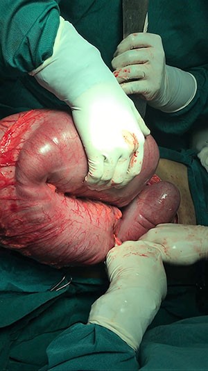
The treatment options for Hirschsprung disease in adults are similar to that in infancy. Duhamel pull through procedure is the surgery of choice.4,11 In cases of skip segmental Hirschsprung disease, it is advisable to take biopsies from multiple sites of proximal normal appearing bowel. Zonal colonic aganglionosis can be managed by segmental resection of the involved segment with primary anastomosis; however, it is important to take biopsies from the rectum to avoid persistence of distal unidentified aganglionic segments, in case of double zonal colonic aganglionosis with a skip segment in between. In this case, left hemicolectomy with primary colorectal anastomosis was done with good short-term outcome. The patient needs long-term follow-up for early diagnosis of recurrent obstruction or persistence of the disease.
Conclusion
Hirschsprung disease should be considered a differential diagnosis in young adults with chronic constipation. The zonal aganglionosis is an atypical presentation of the disease and is extremely rare in the adult population. Thorough evaluation of such patients is necessary, as atypical variants of the disease can be missed on rectal biopsies.
Lessons Learned
Thorough evaluation of young adults with chronic constipation is necessary, as atypical variants can be missed on rectal biopsies. Appropriate surgery can relieve such patients from chronic constipation and prevents major complications.
Authors
Srinivas MVNa, Satyam Gb, Hota PKc, Suhas Md, Jadhav Ve
Correspondence Author
Mallineni Venkata Naga Srinivas
Address: 31-12-3/1, dbrk street
Machavaram, Vijayawada
AP-520004, India
91-9492939357
Vasu.mallineni@gmail.com
Author Affiliations
- Mallineni Venkata Naga Srinivas, MS
Assistant professor
Department of surgery
Mamata general hospital
Khammam, India
- Satyam Guntupalli, MS
Associate professor
Department of surgery
Mamata general hospital
Khammam, India
- Prasan Kumar Hota, MS
Professor & HOD
Department of surgery
Mamata general hospital
Khammam, India
- Suhas Malineni, MBBS
Resident
Department of surgery
Mamata general hospital
Khammam, India
- Varsha Jadhav, MD
Professor & HOD
Department of pathology
Mamata general hospital
Khammam, India
References
- Chen F, Winston JH 3rd, Jain SK, Frankel WL.Hirschsprung's disease in a young adult: report of a case and review of the literature. Ann Diagn Pathol. 2006 Dec;10(6):347-51.
- Fairgrieve J. Hirschsprung disease in the adult. Br J Surg. 1963;50:506-14
- Rosin JD, Bargen JA, Waugh JM. Congenital megacolon of a man 54 years of age: report of case. Proc Staff Meet Mayo Clin. 1950 Dec 20;25(26):710-5.
- Miyamoto M, Egami K, Maeda S et al. Hirschsprung disease in adults: report of a case and review of literature. J Nippon Med Sch. 2005; 72(2):113-120
- O’Donnell AM, Puri P. Skip segment Hirschsprung disease: a systematic review. Paediatr Surg Int. 2010 Nov;26(11):1065-69
- Tiffin MD, Chandler LR, Faber HK. Localised absence of the ganglion cells of the myenteric plexus in congenital megacolon. Am J Dis Child. 1940;59:1071-82
- Moriya H, Naka Zaki H, Yokoyama S et al. A case of segmental aganglionosis localised to descending colon. Jpn J Gastroenterol. 1996;93:39-44
- CG Fu, T Muto, T Masaki, H Nagawa. Zonal adult Hirschsprung disease. Gut. 1996;39: 765-67
- Yeon SK, Joon SL, Kyung Mk et al. Case report: a case of zonal adult Hirschsprung disease. Kor J Neurogastroenterol Motil. 2006;12(2):170-76
- Radu NB, Laura B, Andrea AM et al. Segmental aganglionosis in Hirschsprung disease in newborns: a case report. Rom J Morphol Embryol. 2015;56(2):533-36
- Vorobyov GI, Achkosav SI, Biryukov OM. Clinical features, diagnostics and treatment of Hirschsprung disease in adults. Colorectal Dis. 2010 Dec;12(12):1242-48