Unsupported Browser
The American College of Surgeons website is not compatible with Internet Explorer 11, IE 11. For the best experience please update your browser.
Menu
Become a member and receive career-enhancing benefits

Our top priority is providing value to members. Your Member Services team is here to ensure you maximize your ACS member benefits, participate in College activities, and engage with your ACS colleagues. It's all here.

Become a Member
Become a member and receive career-enhancing benefits

Our top priority is providing value to members. Your Member Services team is here to ensure you maximize your ACS member benefits, participate in College activities, and engage with your ACS colleagues. It's all here.

Become a Member
ACS
Case Reviews in Surgery

Gastrosplenic Fistula in the Setting of Diffuse Large B-Cell Lymphoma Being Treated with Maintenance Chemotherapy

Gastrosplenic Fistula in the Setting of Diffuse Large B-Cell Lymphoma Being Treated with Maintenance Chemotherapy

March 1, 2021

Abstract

Background

Gastrosplenic fistula (GSF) secondary to gastric or splenic lymphoma is a rare event that occurs both primarily in the setting of tumor necrosis and secondary to chemotherapy.

Summary

We report a case of GSF in the setting of diffuse large B-Cell lymphoma (DLBCL) being managed with maintenance chemotherapy. The patient presented with worsening chronic dyspnea, and on computerized tomography (CT) scan to rule out pulmonary embolism, was found to have a large left pulmonary effusion and concern for GSF. Oral contrast administered during a subsequent CT scan of the abdomen and pelvis was seen extravasating into the spleen through a fistulous connection. The patient underwent open splenectomy and partial gastrectomy with an uneventful postoperative course.

Conclusion

GSFs are challenging due to their rarity and propensity to involve surrounding structures. Presentation, diagnosis, and clinical management of this rare complication, as well as a review of the existing literature, are discussed.

Key Words

Gastrosplenic fistula, lymphoma, necrosis, chemotherapy, resection


Case Description

Gastrosplenic fistula (GSF) is a rare but previously described complication of lymphoma. GSF can occur either primarily in the setting of spontaneous tumor necrosis or secondary to chemotherapy induced necrosis. Presentation typically, but not always, involves left upper quadrant pain as well as constitutional symptoms. In some instances, massive hematemesis can occur. Diagnosis and management calls for the surgeon to head a multidisciplinary team comprised of radiologists, gastroenterologists, and oncologists (as is often necessary in complex rare disease-presentations).

This case occurred in the context of a diffuse large B-Cell lymphoma (DLBCL) being managed with chemotherapy. Of the 26 other reports of GSF secondary to lymphoma found in the literature, nine fistulas were discovered after chemotherapy initiation, suggesting that this subset of disease complication presents less frequently than do primary GSFs not undergoing treatment.

A 71-year-old female with a past medical history of non-Hodgkin lymphoma (initially type unknown) underwent six cycles of rituximab, vincristine, cyclophosphamide, and prednisone (RCV-P) followed by maintenance rituximab. Subsequent serial imaging demonstrated potential recurrence of lymphomatous disease characterized by increasing size of an anterior splenic mass. The patient refused further chemotherapy due to poor past tolerance, and she was additionally placed on ibrutinib, which was discontinued after two months due to grade three toxicity, a neutropenic fever. At time of presentation the patient was 17 months out from last induction chemotherapy dose, six months from last maintenance dose, and three months from last ibrutinib dose.

The patient presented to our emergency department with chronic complaints of worsening exertional dyspnea, fatigue, and anorexia. The patient denied abdominal pain, hematemesis, melena, fever, or weight loss. She was tachycardic to 129 bpm but had otherwise normal vital signs. On physical examination, she appeared mildly cachectic, had a minimally distended abdomen, and palpable splenomegaly. No lymphadenopathy was appreciated. Her laboratory values demonstrated a hemoglobin of 8.4 g/dL, which was consistent with her chronic anemia, but were otherwise within normal limits.

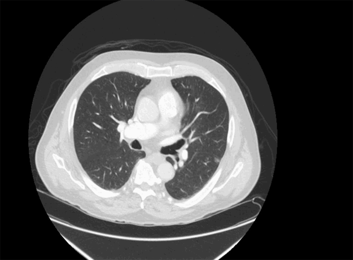
A spiral computerized tomography (CT) scan of the chest was initially performed to rule out pulmonary embolism. Although none was seen, an incidental large left pleural effusion was visualized. Additionally, there was concern for fistulization into the greater curvature of the stomach from the previously followed splenic mass (Figure 1).

Figure 1. Axial imaging demonstrating oral contrast passing between the stomach and the splenic mass through a fistulous tract


Figure 1. Axial imaging demonstrating oral contrast passing between the stomach and the splenic mass through a fistulous tract

A CT of the abdomen and pelvis with oral and intravenous contrast demonstrated interval enlargement of the splenic mass from 9.7x6.7cm six months prior to 11.5x7.6 cm, with heterogeneous areas of air-containing parenchyma indicating necrosis (Figure 2).

Figure 2. Large 11.5x7.6cm splenic mass with heterogeneous areas of air-containing parenchyma indicating necrosis


Figure 2. Large 11.5x7.6cm splenic mass with heterogeneous areas of air-containing parenchyma indicating necrosis

The anterior surface of the mass appeared to invade the greater curvature of the stomach, with ingested oral contrast extending through a fistulous tract between the two. Additionally, the mass directly abutted the left hemidiaphragm, the transverse colon, and the tail of the pancreas.

After undergoing drainage of her exudative pulmonary effusion, assisting her chief complaint of dyspnea, our patient was medically optimized for a radical surgical resection with total parental nutrition, levofloxacin due to concern for pneumonia, and enemas to lessen stool burden in case of possible colonic resection. Through an upper midline incision, exploration confirmed the splenic mass to be invading both the greater curvature of the stomach as well as the left hemidiaphragm. The mass was freed off the diaphragm by blunt dissection, followed by mobilization of the splenic flexure of the colon and spleen. The greater curvature of the stomach and lesser sac were exposed, and no invasion posteriorly into the pancreas was identified. The stomach was then partially resected along the greater curvature using multiple firings of a gastrointestinal anastomosis (GIA) stapler with thick load staples. The splenic artery and vein were dissected and individually transected with a GIA stapler using vascular staples, completing the resection (Figure 3).

Figure 3. Resected gastric greater curvature and spleen


Figure 3. Resected gastric greater curvature and spleen

An intraoperative upper flexible endoscopy demonstrated no bleeding or evidence of leak upon insufflation. Jackson-Pratt drains were left near the gastric staple line and pancreatic tail.
Final pathology demonstrated DLBCL negative for MYC rearrangement, indicating a more favorable prognosis. Appropriate vaccinations were administered postoperatively. After an uneventful recovery period, the patient was discharged on postoperative day seven after removal of her drains, tolerance of a low-residue diet, and with appropriate oncology follow-up to discuss restarting maintenance chemotherapy.

Discussion

It is postulated that GSF formation in lymphoma patients occurs either primarily after spontaneous tumor necrosis23, or secondary to chemotherapy induced cancer cell death. Conceptually, the tumor must breach the splenic capsule and invade the gastric mucosa. Typical presenting features include left upper quadrant pain, fever, fatigue, weight loss, a palpable spleen, lymphadenopathy, or gastrointestinal bleeding.25 Our patient is unique in that she exhibited few typical symptoms and complained primarily of exertional dyspnea likely related to her large left pleural effusion. Investigation of GSF progresses according to presenting symptoms. A high-quality CT scan with contrast (IV and oral) often delineates a fistulous connection, and is prudent for operative planning since nearby structures, such as the diaphragm, colon, and pancreas, are also at risk for tumor invasion. Upper flexible endoscopy can confirm the presence of a fistula but is not necessary.

A search of existing literature through PubMed and Ovid was performed using the search terms "gastrosplenic fistula," "splenic fistula," and "lymphoma." After identifying papers appropriate to the topic, 26 case reports of GSF secondary to lymphoma were confirmed.1-25 Nearly all cases were found in the setting of DLBCL. Cases have been described in other settings, including Crohn disease, gastric adenocarcinoma, gastric ulcers, and trauma, but are exceedingly rare.26,27,28,29 Of the 26 cases of GSF and lymphoma in the literature, 17 were primary GSF in chemo naïve patients, while nine were secondary GSF in the context of previously administered chemotherapy.1-9 15 of 17 (88 percent) primary GSF cases and seven of nine (78 percent) of secondary GSF cases were DLBCL related (Figure 4).

Figure 4. Secondary GSF Cases
Endo=endoscopy, CT=computed tomography, Cysto=cystography, abd=abdomen, PET=positron emission tomography, A/P=abdomen and pelvis, LUQP=left upper quadrant pain, const. sx=constitutional symptoms, LAD=lymphadenopathy, WL=weight loss, SOB=shortness of breath, DOE=dyspnea on exertion, Chemo=chemotherapy, p.gast.=partial gastrectomy, splen.=splenectomy, d. panc.=distal pancreatectomy, RT=radiation therapy, LND=lymph node dissection, t. gast.=total gastrectomy, Dz=disease, yr=year, mo=month, w/o=without, f/u=follow-up, DLBCL= Diffuse Large B Cell Lymphoma, PSL= Primary Splenic Lymphoma, PGL=Primary Gastric Lymphoma

Life-threatening hemorrhage is both a theoretical and described risk, with surgical resection offering definitive management14,16,21 While preoperative splenic embolization may theoretically assist with decreasing bleeding complications, we did not encounter this being done in any of the cases. Additionally, a pathologic diagnosis can potentially change subsequent lymphoma management decisions, as it did in our patient with previously unspecified lymphoma. In our review, resection was performed in 22 patients with both primary and secondary GSF.1-11,13-20,22,23,25 All but one were performed through an open incision. The single case of laparoscopic resection reported a favorable outcome and the patient was disease free at one year.10 GSF resection involves splenectomy and partial gastrectomy with additional radical resection depending on degree of surrounding inflammation and adjacent structure involvement. Seven cases in our review also necessitated a distal pancreatectomy.2,9,12,13,16-17 Fifty percent of cases that underwent resection were lost to follow-up.2,3,5,9,13,14,16,19,20,23,25 Of the remaining 11, three cases reported postoperative mortality at 2 to 5 months from subsequent disease progression1,7,22. Four reported long-term survival at three- to six-year follow-up.4,11,17,18 Adjuvant chemotherapy was administered in 10 of 22 surgically managed cases in the setting of active lymphoma.10-15,17,18,20,23 (Figure 5).

Figure 5. Primary GSF Cases
Endo=endoscopy, CT=computed tomography, Cysto=cystography, abd=abdomen, PET=positron emission tomography, A/P=abdomen and pelvis, LUQP=left upper quadrant pain, const. sx=constitutional symptoms, LAD=lymphadenopathy, WL=weight loss, SOB=shortness of breath, DOE=dyspnea on exertion, chemo=chemotherapy, p.gast.=partial gastrectomy, splen.=splenectomy, d. panc.=distal pancreatectomy, RT=radiation therapy, LND=lymph node dissection, t. gast.=total gastrectomy, Dz=disease, yr=year, mo=month, w/o=without, f/u=follow-up, DLBCL= Diffuse Large B Cell Lymphoma, PSL= Primary Splenic Lymphoma, PGL=Primary Gastric Lymphoma

With a paucity of data on individual chemotherapy regimens in the cases found, it is difficult to find even a correlative link between a particular chemotherapy agent or regimen and GSF formation. Our patient had recently received ibrutinib, which was shortly after discontinued due to toxicity (three months prior to presentation). Again, it is difficult to draw any correlation between this and GSF formation.

Chemotherapy as stand-alone management of GSF without resection was employed in three cases. Two cases21,24 cited the reason for nonoperative management as the patients’ poor health and high perioperative risk; the last case did not specify.12 One case reported disease free survival at publication less than a year later.21 Of the other two, one was lost to follow-up, and the second died at two months after diagnosis.12,24 Additionally, a single nonoperatively managed GSF case secondary to mucosal associated lymphoid tissue (MALT) lymphoma was treated solely with Helicobacter pylori triple therapy after the patient declined resection; there was no reported follow-up.8

Conclusion

Gastrosplenic fistulas are rare events that nearly always occur in the setting of lymphoma, and nearly always with DLBCL. They arise either in the setting of primary tumor necrosis or secondary to chemotherapy induced necrosis that has breached the splenic capsule.

This is the most comprehensive collection of GSF cases collected to date. Our case illustrates the high degree of suspicion that must be exercised in this patient population, as our patient presented with uncommon symptoms of GSF. Known splenic masses in the context of lymphoma should undergo follow-up and serial imaging to monitor for radiographic evidence of complications such as GSF.

Surgical resection with or without adjuvant chemotherapy is the definitive management of GSF if the patient can tolerate an operation due to the risk for hemorrhage described above. Poor surgical candidates can undergo chemotherapy as stand-alone treatment.

Lessons Learned

Gastrosplenic fistulas secondary to lymphoma occur after tumor necrosis either primarily or from chemotherapy. Surgical resection with or without adjuvant chemotherapy is the definitive management of GSF if the patient can tolerate an operation.

Authors

Slagle DLa, Jorge JMb, Nagle Ab

Correspondence Author

Alexander P. Nagle, MD, FACS
Northwestern University Department of Surgery
676 N. St. Clair St., Suite 650
Chicago, IL 60611
312-695-0641
a-nagle@northwestern.edu

Author Affiliations

  1. Feinberg School of Medicine, Northwestern Memorial Hospital, Chicago, IL
  2. Department of General Surgery, Northwestern Memorial Hospital, Chicago, IL

References

  1. Aribaş BK, Başkan E, Altinyollar H, Ungül U, Cengiz A, Erdil HF. . Gastrosplenic fistula due to splenic large cell lymphoma diagnosed by percutaneous drainage before surgical treatment. Turk J Gastroenterol 2008;19(1): 69-70.
  2. Bubenik O, Lopez MJ, Greco AO, Kraybill WG, Cherwitz DL. . Gastrosplenic fistula following successful chemotherapy for disseminated histiocytic lymphoma. Cancer 1983;52(6): 994-996.
  3. Gentilli S, Oldani A, Zanni M. . Gastro-splenic fistula as a complication of chemotherapy for large B cell lymphoma. Ann Ital Chir 2016;87.
  4. Hiltunen KM, Airo I, Mattila J, Helve O. . Massively bleeding gastrosplenic fistula following cytostatic chemotherapy of a malignant lymphoma. J Clin Gastroenterol 1991;13(4): 478-481.
  5. Moghazy KM. Gastrosplenic fistula following chemotherapy for lymphoma. Gulf J Oncolog 2008;(3): 64-67.
  6. Palmowski M, Zechmann C, Satzl S, Bartling S, Hallscheidt P. Large gastrosplenic fistula after effective treatment of abdominal diffuse large-B-cell lymphoma. Ann Hematol 2008;87(4): 337-338.
  7. Seib CD, Rocha FG, Hwang DG, Shoji BT. Gastrosplenic fistula from Hodgkin's lymphoma. J Clin Oncol 2009;27(20): e15-17.
  8. Senapati J, Devasia AJ, Sudhakar S, Viswabandya A. Asymptomatic gastrosplenic fistula in a patient with marginal zonal lymphoma transformed to diffuse large B cell lymphoma—a case report and review of literature. AnnHematol 2014;93(9): 1599-1602.
  9. Sousa M, Gomes A, Pignatelli N, Nunes V. Massive gastrointestinal bleeding after chemotherapy for gastric lymphoma. Int J Surg Case Rep 2016;21: 41-43.
  10. Al-Ashgar HI, Khan MQ, Ghamdi AM, Bamehriz FY, Maghfoor I. Gastrosplenic fistula in Hodgkin's lymphoma treated successfully by laparoscopic surgery and chemotherapy. Saudi Med J 2007;28(12): 1898-1900.
  11. Bird MA, Amjadi D, Behrns KE. Primary splenic lymphoma complicated by hematemesis and gastric erosion. South Med J 2002;95(8): 941-942.
  12. Blanchi A., Bour B, Alami O. Spontaneous gastrosplenic fistula revealing high-grade centroblastic lymphoma: endoscopic findings. Gastrointest Endosc 1995;42(6): 587-589.
  13. Choi JE, Chung HJ, Lee HG. Spontaneous gastrosplenic fistula: a rare complication of splenic diffuse large cell lymphoma. Abdom Imaging 2002;27(6): 728-730.
  14. Dellaportas D, Vezakis A, Fragulidis G, Tasoulis M, Karamitopoulou E, Polydorou A. Gastrosplenic fistula secondary to lymphoma, manifesting as upper gastrointestinal bleeding. Endoscopy 2011;43 Suppl 2 UCTN: E395.
  15. Ding YL, Wang SY. Gastrosplenic fistula due to splenic large B-cell lymphoma. J Res Med Sci 2012;17(8): 805-807.
  16. Favre Rizzo J., López-Tomassetti Fernández E, Ceballos Esparragón J, Santana Cabrera L, Hernández Hernández JR. Massive upper gastrointestinal bleeding secondary to gastrosplenic fistula. Rev Esp Enferm Dig 2013;105(9): 570-871.
  17. Garcia Marin A, Bernardos García L, Vaquero Rodríguez A, Menchén Viso L, Turégano Fuentes F. Spontaneous gastrosplenic fistula secondary to primary gastric lymphoma. Rev Esp Enferm Dig 2009;101(1): 76-78.
  18. Harris NL, Aisenberg AC, Meyer JE, Ellman L, Elman A. Diffuse large cell (histiocytic) lymphoma of the spleen. Clinical and pathologic characteristics of ten cases. Cancer 1984;54(11): 2460-2467.
  19. Jain V, Pauli E, Sharzehi K, Moyer M. Spontaneous gastrosplenic fistula secondary to diffuse large B-cell lymphoma. Gastrointest Endosc 2011;73(3): 608-609.
  20. Kerem M., Sakrak O, Yilmaz TU, Gultekin FA, Dursun A, Bedirli A. Spontaneous gastrosplenic fistula in primary gastric lymphoma: Surgical management. Asian J Surg 2006;29(4): 287-290.
  21. Khan F, Vessal S, McKimm E, D'Souza R. Spontaneous gastrosplenic fistula secondary to primary splenic lymphoma. BMJ Case Rep 2010.
  22. Maillo C Bau J. Gastrosplenic and thoracosplenic fistula due to primary untreated splenic lymphoma. Rev Esp Enferm Dig 2009;101(3): 222-223.
  23. Moran M, et al. Spontaneous Gastrosplenic Fistula Resulting From Primary Gastric Lymphoma: Case Report and Review of the Literature. Balkan Med J 2011;(28): 205-208.
  24. Puppala S, et al. Spontaneous gastrosplenic fistula in primary gastric lymphoma: case report and review of literature. Clinical Radiology Extra 2005;60(2): 20-22.
  25. Rothermel LD, Chadwick CL, Thambi-Pillai T. Gastrosplenic fistula: etiologies, diagnostic studies, and surgical management. Int Surg 2010;95(3): 270-272.
  26. Cary ER, Tremaine WJ, Banks PM, Nagorney DM. Isolated Crohn's disease of the stomach. Mayo Clin Proc 1989;64(7): 776-779.
  27. Krause R, Larsen CR, Scholz FJ. Gastrosplenic fistula: complication of adenocarcinoma of stomach. Comput Med Imaging Graph 1990;14(4): 273-276.
  28. Martinez JD, Moya L, Hernández G, Viola L. Gastrosplenic fistula secondary to gastric adenocarcinoma]. Rev Gastroenterol Peru 2015;35(2): 165-167.
  29. Pizzirusso F, Gillet JP, Fobe D. Isolated spleen metastatic involvement from a colorectal adenocarcinoma complicated with a gastrosplenic fistula. A case report and literature review. Acta Chir Belg 2004;104(2): 214-216.