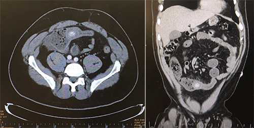
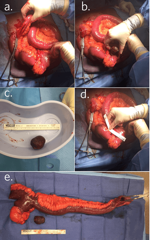
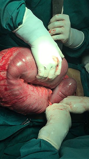
Figure 3. Illustration from Field, et. al.11 (1959) depicting abdominal findings at the time of exploratory laparotomy in a patient with intestinal obstruction due to fecalith formed from a Meckel’s diverticulum. This illustration bears a striking resemblance to the findings in our patient. Reproduced with permission.
Our review of the literature included 10 cases spanning over 50 years. The mean age for presentation was 64 years old with almost equal distribution between male and female patients (6 male:4 female ratio). Males tended to be symptomatic more frequently than female patients.4
The incidence of enteroliths is found to be about 0.3–10 percent of all cases of Meckel diverticulum, with a four percent complication rate. Intestinal obstruction and diverticulitis are the main complications seen in adults, occurring in 40 percent and 20 percent of patients, respectively.1 The incidence of enteroliths formed in a Meckel diverticulum is even lower in children. There was only one case of pediatric small bowel obstruction caused by Meckel enterolith noted in our review of the literature (Table 1).5
Table 1. Review of the literature. (“--“ indicates that this finding was not documented in the literature)
| Case | Author (Year) | Age (years) | Sex | Pre-Operative Imaging Modality and Impression | Pre-operative Diagnosis | Foreign body size | Ectopic Gastric Tissue on Pathology | Surgical Management |
| 1 | Field (1959)11 | 52 | M | Erect x-ray: marked distention of the small bowel, absence of gas in the large bowel. Fluid levels in the small bowel. | Distal small bowel obstruction | “golf-ball-sized” | -- | Fecalith manipulated proximally to Meckel diverticulum. Diverticulectomy was performed. |
| 2 | Bergland (1962)12 | 73 | F | Abdominal x-ray: Distended small intestinal loops with multi-level fluid and gas-filled segments. No free peritoneal air | Diverticulitis of the sigmoid colon with secondary partial small bowel obstruction | 3.0 x 3.0 cm | None | The enterolith was pushed back and removed from the lumen of the distal ileum. The proximal ileum was decompressed by suction. Diverticulectomy and anastomosis were performed. |
| 3 | Christiansen (1967)8 | 48 | F | Abdominal x-ray: Multiple distended loops of small bowel without evidence of colon gas. Laminated opacity in the right lower quadrant.
Decubitus x-ray: multiple varying air fluid levels within the small bowel, and no change in position of the laminated calcification. | Small bowel obstruction with possible gallstone ileus | 3.5 cm | None | Diverticulectomy including enterolith. |
| 4 | Sharma (1970)3 | 68 | F | GI series reported as normal. | -- | Multiple small stones | -- | Diverticulectomy |
| 5 | Benhamou (1979)13 | 78 | M | Abdominal x-ray: Small bowel obstruction with opacity in the right iliac fossa. No radiological evidence of air in the common bile duct. | Gallstone Ileus | -- | None | Enterotomy opposite the diverticulum for stone extraction and diverticulectomy. |
| 6 | Lopez (1991)14 | 85 | M | Supine abdominal x-ray: 3-cm calcified opacity in the right lower quadrant and multiple dilated loops of small bowel | Gallstone Ileus | 3 cm | -- | The enterolith was found within the Meckel diverticulum. Diverticulectomy to remove diverticulum and enterolith was performed. |
| 7 | Lopez (1991)13 | 70 | M | CT: Small bowel obstruction with a foreign body and possible inflammatory focus in the right lower quadrant.
Barium enema: diverticulosis of the sigmoid colon without evidence of diverticulitis. | Small bowel obstruction of unknown etiology | 3.5 cm | -- | The enterolith was mobilized to the more distended small bowel, and a longitudinal enterotomy was performed, and the enterolith was removed. |
| 8 | Rudge (1992)7 | 78 | M | Abdominal x-ray: markedly distended loops of small bowel | Distal small bowel obstruction of unknown etiology | 3.5 x 5cm | None | Diverticulectomy including stone. |
| 9 | Hayee (2003)15 | 79 | F | Abdominal x-ray: opacity on the left side
Gastrografin study: numerous small bowel diverticula of varying sizes and minimal passage of barium beyond the mid-jejunum. | -- | 3 x 4 cm | | The stone was found impacted in the middle of the jejunum and was removed via a small enterotomy. No diverticulectomy was documented. |
| 10 | Lai (2010)4 | 9 | M | Abdominal x-ray: local ileus, multiple dilated bowel loops | Mechanical Ileus | -- | -- | Fecalith was manipulated distally to the cecum, diverticulectomy and appendectomy were performed. |
The cause of enterolith formation is attributed to multiple factors. Poor coordination of the peristaltic wave at the site of the Meckel diverticulum, which produces decreased motility, and slow flow in that area may lead to stone formation.4,6 Meckel enterolith formation is also related to stasis, foreign bodies (such as seeds) acting as a nidus for precipitation of calcium salts.7 The alkaline environment of the Meckel diverticulum, with the absence of ectopic gastric mucosa, leads to precipitation of the enterolith. An alkaline environment favors precipitation of calcium and other minerals from the small intestine, promoting formation of enteroliths.4,8 In the review of the literature, all the pathology from the Meckel diverticula noted that there was no gastric mucosa in the diverticulum. The Meckel enteroliths in our review, when the size was documented, were all at least 3 cm in size.
The Meckel stone ileus abdominal plain film imaging finding is a laminated calcification in the right lower quadrant in the presence of small bowel obstruction.9 Meckel enteroliths are often not well visualized on abdominal radiographs. Ultrasound and abdominal CT scans are more sensitive imaging modalities for diagnosing an enterolith.10 However, the diagnosis is typically made at the time of surgery.5 No definitive diagnosis was made on imaging prior to surgery in our case report review.
A Meckel Scan, a technetium pertechnetate scan, has been used to detect Meckel diverticula since 1970. The technetium pertechnetate relies on the presence of at least 1.8 cm2 gastric mucosa in the diverticula. In our review, there was no gastric mucosa identified on any pathology of the Meckel diverticula rendering this modality ineffective at diagnosing a diverticula generating a Meckel stone ileus.2
The surgical management of a fecalith generated from a Meckel diverticulum causing small bowel obstruction varied in our review of the literature. In the 10 cases reviewed, three reports described performing an enterotomy to remove the Meckel stone ileus. Stone manipulation more proximal to the site of the Meckel diverticulum and more dilated bowel was described in three cases prior to resection. All case reports the Meckel diverticulum was removed. The pediatric case described a simultaneous appendectomy. The surgical management of the cases is summarized in Table 1.
According to DiGiacomo, if a pathologic Meckel diverticulum is encountered at laparotomy with evidence of chronic or previous inflammation, the diverticulum should be removed. The treatment of choice is ileal resection for Meckel diverticula. Although simple excision of the diverticula may cause less morbidity, there is the possibility of ectopic gastric or pancreatic tissue extending beyond the mouth of the diverticula, which cannot be determined by palpation and necessitates removal of the entire segment of bowel with the diverticulum.
10Conclusion
Meckel stone ileus should be considered preoperatively in a patient who presents with a small bowel obstruction secondary to a foreign body 3 cm or greater on imaging in the distal small bowel with an absence of gallbladder pathology. All cases reviewed have documented the absence of gastric mucosa on pathology specimens in the Meckel diverticulum generating a fecalith. This indicates the role of an alkaline environment in the diverticulum combined with poor peristalsis and foreign bodies as a nidus for the formation of the fecalith. Additionally, it indicates that the Meckel scan would not visualize a stone generating Meckel diverticula. Surgical management varies in the literature review; in each case the Meckel diverticulum was removed with either an enterotomy, diverticulectomy or removing the entire segment of bowel. Current surgical management is to remove the entire segment of bowel adjacent to the diverticula to remove all ectopic mucosa.2 The fecalith can be addressed surgically by creating an enterotomy to remove the fecalith at the site of impaction, manipulating the stone distally to the cecum, or mobilizing the stone proximally into the diverticula before resection to minimize the amount of bowel resected.
Lessons Learned
Meckel diverticula can present with complications well past adolescence. Meckel stone ileus should be considered preoperatively in patients who present with small bowel obstruction secondary to a foreign body 3 cm or greater on imaging in the distal small bowel with an absence of gallbladder pathology.
Authors
Stutz KN, Blackwell LM
Correspondence Author
Kathleen N. Stutz c/o Dr. Lea Blackwell
Associates in General Surgery
21 Barkley Circle
Fort Myers, FL 33907
425-445-0494
kathleen.stutz@gmail.com
Author Affiliations
Healthpark Medical Center
Department of Surgery
Fort Myers, FL
References
- Bennett GL, Birnbaum BA, Balthazar EJ. CT of Meckel Diverticulitis in 11 Patients. Am J Roentgenol. 2004;182: 625-29.
- Javid P, Pauli EM. Meckel Diverticulum. UpToDate. N.p., 11 May 2016.
- Sharma G, Benson CK. Enteroliths in Meckel Diverticulum: Report of a Case and Review of the Literature. Can J Surg. 1970;13: 54-58.
- DiGiacomo JC, Cottone FJ. Surgical Treatment of Meckel Diverticulum. South Med J. 1993;86(6):671-5.
- Lai HC. Intestinal Obstruction Due to Meckel Enterolith Pediatr Neonatol. 2010;51(2):139-40.
- Frazzini VI, English WJ, Bashist B, Moore E. Case Report. Small Bowel Obstruction Due to Phytobezoar Formation within Meckel Diverticulum: CT Findings. J Comput Assist Tomogr. 1996;20(3):390-2
- Macari M, Panicek DM. CT Findings in Acute Necrotizing Meckel Diverticulitis Due to Obstructing Enterolith. J Comput Assist Tomogr. 1995;19(5):808-10.
- Rudge FW. Meckel Stone Ileus. Mil Med. 1992;157(2):98-100..
- Christiansen KH, Cancelmo RP. Meckel Stone Ileus. Am J Roentgenol Radium Ther Nucl Med. 1967;99(1):139-41.
- Pantongrag-Brown L, Levine MS, Buetow PC, Buck JL, Elsayed AM. Meckel Enteroliths: Clinical, Radiologic, and Pathologic Findings. AJR Am J Roentgenol. 1996;167(6):1447-50.
- Field RJ Sr., Field RJ Jr. Intestinal Obstruction Produced by Fecalith Arising in Meckel Diverticulum. AMA Arch Surg. 1959;79(1):8-9.
- Bergland RM, Gump F, Price JB Jr.. An Unusual Complication of Meckel Diverticula Seen in Older Patients. Ann Surg. 1963;158:6-8.
- Benhamou, G. Small Intestinal Obstruction by an Enterolith from a Meckel Diverticulum. Int Surg. 1979;64(1):43-5.
- Lopez P V, Welch JP. Enterolith Intestinal Obstruction Owing to Acquired and Congenital Diverticulosis. Dis Colon Rectum. 1991;34(10):941-4.
- Hayee B, Kahn HN, Al-Mishlab T, McPartlin JF. A Case of Enterolith Small Bowel Obstruction and Jejunal Diverticulosis. World J Gastroenterol. 2003;9(4):883-4.

