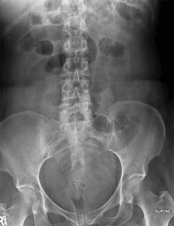
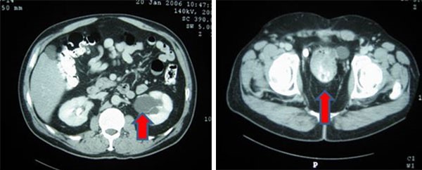
Laboratory work-up for systemic disease, including CBC, CMP, hepatitis panel, ANA, ANCA, SSA/SSB, lupus anticoagulant, anticardiolipin antibodies, and anti-beta 2 glycoprotein antibodies, was negative. Chest x-ray revealed no abnormalities. Colonoscopy was negative for inflammatory bowel disease and malignancy.
Discussion
Pyoderma gangrenosum is a rare but serious post-operative complication. Diagnosis and treatment is often delayed due to confusion with surgical site infection.2,11,12 PG should be considered in the differential diagnosis for all early post-operative skin changes, especially when lesions are progressive despite appropriate antibiotic therapy, exacerbated by surgical debridement, and in the setting of systemic disease (inflammatory bowel disease, hematologic disorders, autoimmune syndromes or malignancy).
Diagnosis is based on characteristic clinical and pathologic features. The hallmark lesion of classical PG is a painful, rapidly enlarging, violaceous ulcer with overhanging or undermined borders and a necrotic base.13 Histopathology nearly always reveals neutrophilic dermal infiltration. Leukocytoclasia, abscess formation, and leukocytoclastic vasculitis are less frequently observed. Although the pathological findings are not specific, biopsy is important to rule out other causes of ulceration such as infection, vasculitis, or malignancy.14
PG is associated with systemic disease in 33 to 84 percent of cases.14 Inflammatory bowel disease is most frequent coincident pathology (20 to 30 percent).14 Other associations include hematologic disorders (monoclonal gammopathy, myelofibrosis, myelogenous leukemia, hairy cell leukemia), autoimmune disease, solid tumors, and infection (HIV, chronic hepatitis). Skin lesions can occur before, after, or coincident with systemic illness. Additional work-up should include laboratory studies (CBC, ESR, liver and kidney profiles, serum and urine protein electrophoresis, coagulation panel, antiphospholipid antibodies, ANA, cryoglobulins), peripheral blood smear, bone marrow aspirate, colonoscopy, and chest X ray.2 Treatment of the underlying condition is critical.
Treatment of pyoderma skin lesions consists of local and systemic therapy. Local wound care focuses on maintaining optimum moisture balance in the lesion. Topical medications commonly used include corticosteroids, tacrolimus,15 and sodium cromoglycate. Intralesional injections of corticosteroids, cyclosporine,16 and phenytoin17 have also been effective. Systemic therapy should be considered for aggressive lesions. Corticosteroids and cyclosporine are first line treatments. Immunomodulatory therapy and biologic response modifiers, such as mycophenolate mofetil, tacrolimus, dapsone, azathioprine, and infliximab, are also sometimes used.14
Surgery should be avoided on active PG lesions. Debridement may exacerbate the problem due to a pathergic response, in which minor skin trauma results in additional ulceration. If surgery is unavoidable, it should be performed only after inflammation has been controlled with topical and systemic immunosuppresants.13
Effective management of soft tissue defects associated with PG is a challenge. Numerous reconstructive options normally exist for large, ulcerative lesions, including healing by secondary intention, delayed primary wound closure, skin grafting, local or regional flaps, and free tissue transfer18; however, extensive surgery should be avoided in patients with a history of PG for the above reasons. Split thickness skin grafting has been used with some success but recurrence of PG at the recipient site has been reported in several cases.13 Donor site morbidity is also a concern as new PG lesions may develop at these locations as well. Amputation is a last resort but is known to improve quality of life in patients with severe, refractory PG.19
External tissue expanders have been used to reduce wound burden in traumatic soft tissue defects,20 fasciotomy wounds,21 donor sites after flap harvest,22 and wounds resulting from oncologic resection.23,24 Mechanical manipulation of the skin has been utilized since the 1950s25 and is based on the principals of biological creep, mechanical creep, and stress relaxation. When skin is stretched beyond its physiologic limit, transmembrane mechanoreceptors induce a series of events that result in increased mitotic activity and collagen synthesis, a process termed biological creep. The effect is increased tissue mass. Existing collagen fibers elongate and realign parallel to each other, allowing the skin to stretch. The increase in length of a tissue mass is called mechanical creep. Stress relaxation is defined as the decrease in retractive force exhibited by a material when it is held at a given stretch over time. The end result is an increase in skin surface area when an external force is applied over time.26
External tissue expanders facilitate closure of full-thickness defects by applying continuous traction on the skin. A number of methods can be used for external expansion, including fashioning vessel loops into a Jacob’s ladder,21 rubber banding,27 and retention sutures.28 An off-the-shelf device (DermaClose® RC, Wound Care Technologies, Inc, Chanhassen Minnesota) is now available to simplify the process of application and control the amount of force applied to the wound edges (Figure 6). Stainless steel anchors are placed in healthy skin 1–3 cm from wound edge and 2–3 cm apart from each other and secured with surgical staples. A nylon monofilament is laced through the anchors and force is applied by a tension controller. The tension controller is spring activated and internally calibrated such that once the device is activated, no additional adjustments are necessary. A constant force is applied to the wound edges while the device is in place.29
There are many advantages of external tissue expansion over other reconstructive methods, particularly in the treatment of PG. The technique is noninvasive, causes minimal trauma to the skin and has no donor site morbidity. Expansion creates phenotypically similar skin that is matched tissue in color, texture, thickness, and hair bearing status, leading to excellent cosmesis.24 The device is easy to apply and use, achieves wound closure (or significantly reduces wound burden) in a short period of time,20 and is cost effective.30,31 In addition, external tissue expanders may be used in conjunction with NPWT to decontaminate and achieve wound closure simultaneously.20
Conclusion
Effective management of postoperative PG is a challenge. The authors report the safe, effective use of an external tissue expansion device as a possible treatment option for the management of soft tissue defects associated with PG. Further studies are needed to validate applications for external tissue expanders.
Lessons Learned
Known complications of external tissue expansion include blistering or maceration of the skin, peri-wound tissue ischemia or necrosis,22 and scarring. These can be avoided by careful device positioning and protection (e.g., placing soft foam dressings underneath the skin anchors and tension controller),20 close monitoring of surrounding tissue,22 and removal of the device after a maximum of seven days.29
Authors
Sarah E. Sasor, MD
Indiana University School of Medicine
Indianapolis, IN
Julia A. Cook , MD
Indiana University School of Medicine
Indianapolis, IN
Michael W. Chu, MD
Kaiser Permanente
Los Angeles, CA
William A. Wooden, MD, FACS
Indiana University School of Medicine
R.L. Roudebush VA Medical Center
Indianapolis, IN
Sunil Tholpady, MD, PhD, FACS
Indiana University School of Medicine
R.L. Roudebush VA Medical Center
Indianapolis, IN
Juan Socas, MD
Indiana University School of Medicine
Indianapolis, IN
Correspondence
Sarah E. Sasor, MD
Division of Plastic Surgery
Indiana University School of Medicine
545 Barnhill Drive
EH 232
Indianapolis, IN 46202
Phone: 908-892-7745
Fax: 317-278-87465
E-mail: ssasor@gmail.com
Disclosures
The authors have no conflicts of interest to disclose.
References
- de Thomasson E, Caux I. Pyoderma gangrenosum following an orthopedic surgical procedure. Orthop Traumatol Surg Res. 2010;96(5):600-602.
- Wanich T, Swanson AN, Wyatt AJ, et al. Pyoderma gangrenosum following patellar tendon repair: a case report and review of the literature. Am J Orthop 2012;41(1):E4-9.
- Carrasco Lopez C, Lopez Martinez A, Roca Mas JO, et al. Pyoderma gangrenosum in breast reconstruction. J Plast Surg Hand Surg. 2012;46(3-4):281-282.
- Caterson SA, Nyame T, Phung T, et al. Pyoderma gangrenosum following bilateral deep inferior epigastric perforator flap breast reconstruction. J Reconstr Microsurg. 2010;26(7):475-479.
- Rietjens M, Cuccia G, Brenelli F, et al. A pyoderma gangrenosum following breast reconstruction: a rare cause of skin necrosis. Breast J. 2010;16(2):200-202.
- Rajapakse Y, Bunker CB, Ghattaura A, et al. Case report: pyoderma gangrenosum following Deep Inferior Epigastric Perforator (Diep) free flap breast reconstruction. J Plast Reconstr Aesthet Surg. 2010;63(4):e395-396.
- MacKenzie D, Moiemen N, Frame JD. Pyoderma gangrenosum following breast reconstruction. Br J Plast Surg. 2000;53(5):441-443.
- Armstrong PM, Ilyas I, Pandey R, et al. Pyoderma gangrenosum. A diagnosis not to be missed. J Bone Joint Surg Br. 1999;81(5):893-894.
- Nakajima N, Ikeuchi M, Izumi M, et al. Successful treatment of wound breakdown caused by pyoderma gangrenosum after total knee arthroplasty. Knee. 2011;18(6):453-455.
- Schoemann MB, Zenn MR. Pyoderma gangrenosum following free transverse rectus abdominis myocutaneous breast reconstruction: a case report. Ann Plast Surg. 2010;64(2):151-154.
- Ayestaray B, Dudrap E, Chartaux E, et al. Necrotizing pyoderma gangrenosum: an unusual differential diagnosis of necrotizing fasciitis. J Plast Reconstr Aesthet Surg. 2010;63(8):e655-658.
- Williamson KD, Nguyen NQ. A large shin ulcer after minor trauma: please do not debride! Gastroenterol. 2012;143(4):e11-12.
- Ratnagobal S, Sinha S. Pyoderma gangrenosum: guideline for wound practitioners. J Wound Care. 2013;22(2):68-73.
- Pereira N, Brites MM, Goncalo M, et al. Pyoderma gangrenosum - a review of 24 cases observed over 10 years. Int J Dematol. 2013;52(8):938-45.
- Schuppe HC, Homey B, Assmann T, et al. Topical tacrolimus for pyoderma gangrenosum. Lancet. 1998;351(9105):832.
- Mokni M, Phillips TJ. Management of pyoderma gangrenosum. Hosp Pract. 2001;36(4):40-44.
- Fonseka HF, Ekanayake SM, Dissanayake M. Two percent topical phenytoin sodium solution in treating pyoderma gangrenosum: a cohort study. Int Wound J. 2010;7(6):519-523.
- Levin LS. The reconstructive ladder. An orthoplastic approach. Orthop Clin North Am. 1993;24(3):393-409.
- Powell FC, Su WP, Perry HO. Pyoderma gangrenosum: classification and management. J Am Acad Dermatol. 1996;34(3):395-409; quiz 410-392.
- Santiago GF, Bograd B, Basile PL, et al. Soft tissue injury management with a continuous external tissue expander. Ann Plast Surg. 2012;69(4):418-421.
- Fowler JR, Kleiner MT, Das R, et al. Assisted closure of fasciotomy wounds: A descriptive series and caution in patients with vascular injury. Bone Joint Res. 2012;1(3):31-35.
- Durden F, Jr., Tiwari P, Kocak E. Can the DermaClose device contribute to periwound tissue ischemia and necrosis: a case presentation and discussion? Plast Surg Nurs. 2012;32(3):132-133.
- Bajoghli AA, Yoo JY, Faria DT. Utilization of a new tissue expander in the closure of a large Mohs surgical defect. J Drugs Dermatol. 2010;9(2):149-151.
- Laurence VG, Martin JB, Wirth GA. External tissue expanders as adjunct therapy in closing difficult wounds. J Plast Reconstr Aesthet Surg. 2012;65(10):e297-299.
- Neumann CG. The expansion of an area of skin by progressive distention of a subcutaneous balloon; use of the method for securing skin for subtotal reconstruction of the ear. Plast Reconstr Surg (1946). 1957;19(2):124-130.
- Wilhelmi BJ, Blackwell SJ, Mancoll JS, et al. Creep vs. stretch: a review of the viscoelastic properties of skin. Ann Plast Surg. 1998;41(2):215-219.
- Petroianu A. [Synthesis of large wounds of the body wall with rubber elastic band]. Acta Med Port. 2011;24(3):427-430.
- Gruver D. A no. 2 Prolene suture as a tissue expander. Plast Reconstr Surg. 2005;116(1):352.
- Dermaclose. Instructions for Use. 2011. www.dermaclose.com/instructions.pdf. Accessed June 20, 2013.
- O'Reilly AG, Schmitt WR, Roenigk RK,et al. Closure of scalp and forehead defects using external tissue expander. Arch Facial Plast Surg. 2012;14(6):419-422.
- Formby P, Flint J, Gordon WT, et al. Use of a continuous external tissue expander in the conversion of a type IIIB fracture to a type IIIA fracture. Orthopedics. 2013;36(2):e249-251.


