Unsupported Browser
The American College of Surgeons website is not compatible with Internet Explorer 11, IE 11. For the best experience please update your browser.
Menu
Become a member and receive career-enhancing benefits

Our top priority is providing value to members. Your Member Services team is here to ensure you maximize your ACS member benefits, participate in College activities, and engage with your ACS colleagues. It's all here.

Become a Member
Become a member and receive career-enhancing benefits

Our top priority is providing value to members. Your Member Services team is here to ensure you maximize your ACS member benefits, participate in College activities, and engage with your ACS colleagues. It's all here.

Membership Benefits
ACS
Case Reviews in Surgery

Microscopic Submucosal Lipomas with Mucosal Ulceration Can Be a Cause of Intermittent Rectal Bleeding

Colonic lipomas are a rare entity and are reported in the literature with a variety of presentations.

August 29, 2020

Abstract

Background

Colonic lipomas are a rare entity and are reported in the literature with a variety of presentations. Although they can mimic malignant lesions, they are benign proliferative lesions of fatty tissue arising from the submucosa of the intestine. They can present with crampy abdominal pain associated with obstruction, and they most commonly present with intussusception and bowel obstruction. They are also reported as causes of gastrointestinal bleeding. Microscopic submucosal lipomas with mucosal ulceration associated with either chronic anemia and slow gastrointestinal bleeding or intermittent bleeding causing bright red blood per rectum have not been reported in the literature.

Summary

We report a case in which microscopic submucosal lipomas were associated with bright red blood per rectum with an initial presentation of sepsis secondary to colitis. A 55-year-old female admitted with the diagnosis of colitis started having a drop in hemoglobin and bloody bowel movements. A CTA and nuclear scan could not locate the source of bleeding. She had findings of sigmoid and transverse colon colitis on CT scan. Colonoscopy showed sigmoid and ascending colon colitis without any evidence of active bleeding. The patient was taken to the operating room in the setting of bleeding refractory to conservative management, and a right colectomy was performed. The specimen showed scattered ulceration of the ascending colon and a friable terminal ileum.

Conclusion

The patient’s hemoglobin was stable after the surgery, and pathology showed submucosal lipomatosis with overlying mucosal ulcerations. Microscopic submucosal lipomatosis with mucosal ulceration may present as intermittent bleeding. Pathologists should keep this differential in mind because it may lead to differences in management.

Key Words

Colonic lipomas, colonic lipomatosis, submucosal colon lipoma, colon mucosal ulceration, submucosal lipomatosis, bowel lipoma


Colonic lipomas are a rare entity and are reported in the literature with a variety of presentations. In the myriad of a long list of differential diagnoses for bright red blood per rectum, colonic lipomas should have their own place. Although colonic lipomas can mimic malignant lesions, they are benign proliferative lesions of fatty tissue arising from the submucosa of the intestine. Lipomatous disease of the intestine is a rare phenomenon that mostly presents in the colon with right-sided predominance. It is a disease of the older age group, with predominance in the sixth decade of life. Most of the time, these are asymptomatic lesions found incidentally on endoscopy or on CT scan done for other reasons.1 The lesions have been reported as incidental findings upon autopsy, with an incidence of 0.3-0.5 percent.2 The fatty proliferation leads to an appearance of pedunculated polyps, and, when multiple in number, can be hard to differentiate from familial adenomatous polyposis.3 The intraluminal growth and polyp formation mostly causes crampy abdominal pain associated with obstruction, and the condition most commonly presents with intussusception and bowel obstruction.4 This entity that is described in the literature with visible polyposis lipomatosis on colonoscopy is the most common presentation and is most commonly diagnosed on colonoscopy.

There are cases reported in the literature where colonic lipomas cause sigmoid perforation, mechanical obstruction of the transverse colon, and massive intestinal bleeding associated with hematochezia.5-7 A very small proportion of these colonic lipomas are associated with gastrointestinal bleeding. When the lipomas are small, biopsies of these lesions can miss the diagnosis if the pathologist is not looking for submucosal lipomas as part of the differential diagnosis. The lesions may simply be reported as colitis—this is what occurred in our case report.

The microscopic submucosal lipomas associated with mucosal ulceration (with either chronic anemia and slow gastrointestinal bleeding or with intermittent bleeding causing bright red blood per rectum) have not been reported in the literature. Our case involves microscopic submucosal lipomas associated with bright red blood per rectum with an initial presentation of sepsis secondary to colitis.

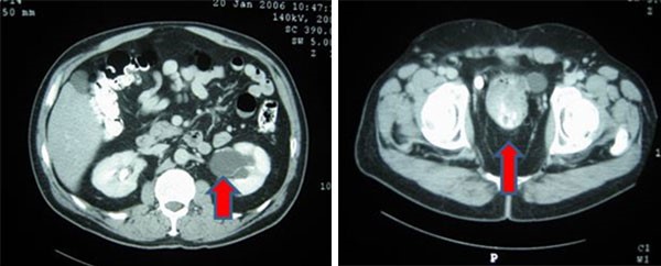
A 55-year-old female presented to the emergency room with bilateral lower abdominal crampy pain associated with two to three episodes of vomiting per day, six to seven episodes of non-bloody diarrhea, and subjective chills for the past four days. She denied any weight loss or any similar episodes in the past. Her last colonoscopy was performed four months prior and showed benign polyps. The patient denied a history of atrial fibrillation and was not on blood thinners. She had a past medical history of hypertension, hyperlipidemia, and chronic kidney disease stage 3. The patient also had a past surgical history of a left donor nephrectomy 10 years ago and laparoscopic cholecystectomy six years ago. She smoked half a pack of cigarettes per day for 25 years. The patient’s mother had end-stage renal disease, with no family history significant for inflammatory bowel disease or colon cancer. On physical exam of her abdomen, the patient had tenderness in the bilateral lower quadrants but no guarding. The patient had a temperature of 39.5°C in the emergency room, a heart rate of 120, and blood pressure= 90/56. Her white blood cell count was 3.0 and hemoglobin was 15.3, with a creatinine of 1.46 and lactic acid of 1.1. A CT scan was obtained, which showed mild sigmoid colitis and proximal to mid transverse colon colitis (Figures 1 and 2). Blood cultures were drawn, and the patient was started on broad spectrum antibiotics and was admitted.

Figure 1. CT scan showing ascending colon wall thickening.

Figure 2. CT scan showing mild sigmoid colitis and wall thickening.

The next day, the patient started having loose bloody bowel movements. Her hemoglobin dropped from 15.3 to 7.8 in the next three days. The patient continued to have intermittent bloody bowel movements but remained hemodynamically stable. A CT angiogram was obtained that showed patent mesenteric vasculature (Figure 3); nuclear bleeding scan did not show any source of bleeding. A colonoscopy was performed that showed external hemorrhoids, 3 cm sigmoid ulceration associated with edema, and ascending colon colitis without evidence of active bleeding. Biopsies of the ascending colon were obtained (Figure 4). Stool studies were negative for Clostridium difficile, stool ova, and parasite and stool toxins. Blood cultures that were drawn on admission grew Enterococcus avium, Steptococcus viridans, and Streptococcus parasanguinus; however the patient remained afebrile after the day of admission, and her white blood cell count remained normal. Biopsies from the colonoscopy showed early colitis.

Figure 3. CT angiogram showing patent mesenteric vasculature.

Figure 4. Colonoscopy showing 3cm sigmoid ulceration associated with edema and ascending colon colitis without evidence of active bleeding.

On hospital day 10, the patient had continued intermittent bloody bowel movements, and her hemoglobin dropped as low as 6.3. The patient received six units of packed red blood cells during her hospitalization to date. The decision was made to take the patient to the operating room with the plan of a right hemicolectomy and end ileostomy, and, if the patient continued to bleed postoperatively, then to proceed with a total abdominal colectomy. Intraoperatively, there were no gross abnormalities seen in any part of colon from the serosal side. The decision was made to perform the right hemicolectomy with end ileostomy. The colon specimen was opened during the case, and showed friable terminal ileum mucosa and some small scattered ulcerations in the ascending colon (Figures 5 and 6). The patient tolerated the procedure well. On postoperative day 4, the patient’s hemoglobin was stable, she tolerated a regular diet, her ostomy was productive, and she was discharged home. Pathology from the specimen showed submucosal lipomatosis with mucosal ulceration of mucosa covering the microscopic submucosal lipoma. There was no evidence of colitis, ischemia, or malignancy (Figure 7).

Figure 5. Intraoperative colon specimen small scattered ulcerations in the ascending colon.

Figure 6. Intraoperative colon specimen showing friable terminal ileum mucosa.

Figure 7. Pathology from the specimen showing submucosal lipomatosis with mucosal ulceration of mucosa covering the microscopic submucosal lipoma.

Discussion

Submucosal lipomas with growth into the lumen causing intussusception usually present with large bowel obstruction. The diagnosis is often made on specimen pathology once the patient has been operated on, as it masquerades as malignancy on CT scan. Patients with intermittent obstruction and CT and endoscopic evidence of a polypoid mass with an inconclusive biopsy of the mass should be further investigated (while considering submucosal lipomas as the entity in mind). Patients with diffuse lipomatosis can have numerous polyps throughout the colon and can be confused with having familial adenomatous polyposis. When attempting to make a diagnosis, submucosal lipomas should be considered as part of the differential diagnosis. Submucosal lipomas are reported as a cause of gastrointestinal bleeding. Frequently, they present as macroscopic lipomas, where those that are <2cm in size can be treated with endoscopic resection, and larger ones can be treated with colon resection.8

Endoscopic treatment of microscopic lipomas are not defined in the literature, as these lesions are not identifiable on colonoscopy, and the stigmata of ulceration, which is usually seen and biopsied in these cases as colitis. The knowledge of the existence of such diagnoses to a pathologist and gastroenterologist can shift the management of this disease towards pursuing endoscopic or surgical resection.

The intermittent bleeding from these small lesions are not picked up by nuclear bleeding scans or CT angiography. The bleeding can also be masked during colonoscopy, since they intermittently slough off the mucosa covering them—which does not shows up as active bleeding on colonoscopy. The mucosal ulcerations associated with submucosal lipomas can cause bacterial translocation and can be responsible for sepsis secondary to bacteremia, as seen in our patient. This further prolongs the diagnosis because patients are presumed to have either infectious or ischemic colitis as the cause of bleeding , the management of which follows a different algorithm.

Conclusion

In summary, the diagnosis for microscopic submucosal lipomatosis with mucosal ulceration is challenging and should be kept in mind when working up a patient with prolonged gastrointestinal bleeding without any identifiable cause; this condition should be managed accordingly.

Lessons Learned

Microscopic submucosal lipomatosis with mucosal ulceration may present as intermittent bleeding. Pathologists should keep this differential in mind because it may lead to differences in management. Smaller macroscopic lipomas <2cm can be treated with endoscopic resection, while larger ones are amenable to colon resection.

Authors

Shaan E Alam, MDa; Timothy Bechtel, MD, FACSb; Benjamin YM Tan, MDa; and Dinesh Vyas, MD, FACSa

Correspondence Author

Dr. Benjamin YM Tan
Department of Surgery
San Joaquin General Hospital
500 W Hospital Rd
French Camp, CA 95231
(209) 468-8820
ymtan@sjgh.org

Author Affiliations

a Department of Surgery, San Joaquin General Hospital
b Department of Surgery, Saint Joseph Medical Center

Disclosure Statement

The authors have no conflicts of interest to disclose.

Acknowledgements

We would like to thank Dr. Lily Pang for her assistance in providing pathology images.

References

  1. Day DW, Jass JR, Price AB, et al.Morson and Dawson’s Gastrointestinal Pathology. 4th Ed. USA: Blackwell Science Ltd; 2008:Chapter 39.
  2. Mandal S. Lipomatous Polyp Presenting With Intestinal Intussusception in Adults: Report of Four Cases. Gastroenterol Res. 2010;3(5):229-231.
  3. Zarrin-Khameh N, Haas EM, Ro J, Thrall MJ. Lipomatosis coli, a mimicker of familial polyposis. Ann Diagn Pathol. 2013;17(2):210-213.
  4. Megaly M, Yacoub G. Transverse Colon Submucosal Lipoma Presenting With Colocolic Intussusception. ACG Case Rep J. 2016;3(3):158-159.
  5. Kumar L, Laksman TK. Giant submucosal lipomatous polyp causing perforation of sigmoid colon: a case report and review of literature. J Clin Diagn Res. 2015;9(1):PD22-4.
  6. Coyne P, Teemul T, Dent B, Henderson D, Crabbe R, Garud T. Submucosal lipoma of the large intestine masquerading as a colonic malignancy. J Surg Case Rep. 2011;2011(2):4.
  7. Vasiliadis K, Katsamakas M, Nikolaidou A, Christoforidis E, Tsalis K, Tsalikidis A. Submucosal Lipoma of the Ascending Colon as a Source of Massive Lower Gastro-intestinal Bleeding: a Case Report. Acta Chir Belg. 2008;108(3):356-359.
  8. De Ruijter SH, van Marle AG, Doornewaard H, Melse JC. Submucosal lipoma of the colon: abdominal cramps with rectal bleeding and weight loss. Ned Tijdschr Geneeskd. 2006;150(36):1990-1993.