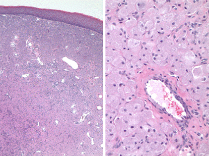
Figure 3. A) H&E stain, 4x. There is a subepithelial proliferation of cells with abundant eosinophilic cytoplasm. Note the absence of hyperplasia of overlying squamous epithelium and the prominence of vascular structures. B) H&E stain, 20x. The tumor cells have abundant granular cytoplasm (low nuclear:cytoplasmic ratio) and small, uniform nuclei.
Discussion
The pathogenesis of congenital epulides is not known. Citing the preferential occurrence in females, some authors suggest the influence of hormones on development.8 Potential cells of origin include fibroblasts, pericytes, epithelial and undifferentiated mesenchymal cells, myofibroblasts, and neuron-related cells.9
The first indication that a CE is present may be on prenatal ultrasound. In utero congenial epulis may lead to polyhydramnios. In the absence of other causes of polyhydramnios, careful evaluation of the oronasal cavity by ultrasound may be useful in the identification of CE or similar tumors of the oral cavity.10,11 When recognized before birth, three-dimensional ultrasound may be useful to evaluate fetal swallowing and airway patency. In utero identification allows for adequate preparation at parturition for the possibility of airway obstruction.6 The differential diagnosis of an anterior oral mass includes congenital epulis, teratoma, odontogenic cyst, dermatofibrosarcoma protuberans, and granular cell tumor.
In order to determine the definitive diagnosis, histologic factors, patient factors, gross morphologic factors, and imaging can assist in discriminating between the different potential differential diagnoses.
Histologically similar to adult granular cell tumors, CEs can be differentiated by negative S-100 staining.9 Absence of cytoplasmic hyaline granules, solid growth pattern, pericytic proliferation, and attenuated overlying epithelium are also key CE discerning characteristics. In our case, microscopy revealed a subepithelial proliferation of cells with abundant eosinophilic cytoplasm in the absence of hyperplasia of overlying squamous epithelium is evident (Figure 3). Prominent vascular structures are typically appreciated as well.
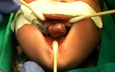
CE can also be distinguished from granular cell tumors by patient characteristics such as age. On gross inspection, a flesh colored, pendulous, and firmly adherent mass to the maxillary alveolar margin is the typical presentation.
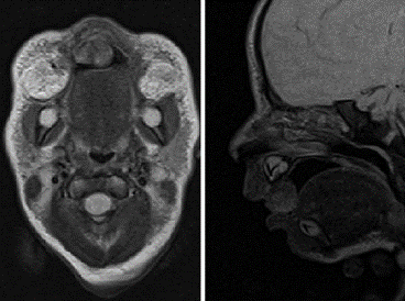
MRI can further assist in confirming a diagnosis of CE. On MRI, a heterogeneous mass on T2 images and an isodense mass on T1 images are seen. Cartilaginous, mucosal, and fibrofatty elements are typically appreciated, while dental elements within the lesion are less common.
Surgical intervention is typically required for the treatment of congenital epulis. Expedited surgery may be required in the case of oral airway obstruction, whereas surgical excision for patients with feeding failure secondary to CE can be performed semi-electively. The size of the CE influences the degree of obstruction and its impact upon successful feeding. Larger lesions are more likely to obstruct the upper airway, leading to dyspnea and possibly hypoxemia. Suffocation from a CE has not been previously reported but is possible if the CE is large enough to obstruct the airway entirely. Although reports of spontaneous regression exist, the potential morbidity and mortality risks warrant surgical excision, especially for large lesions.5,6 Some authors suggest conservative management of small CEs that do not compromising feeding or the neonatal airway as growth halts after birth and spontaneous involution has been reported.12 Because of the small number of reported cases, recurrence rate after surgical excision is unknown.
Close coordination between a pediatric plastic surgeon and a pediatric anesthesiologist is useful in planning perioperative care. Pediatric airways may be challenging, and the presence of a large anterior mass further confounds securing a safe airway in some cases. Airway complications including oral bleeding, aspiration, and pneumothorax may occur, making successful intubation more of a challenge.13,14
Conclusion
In summary, CE is a rare and benign oral cavity lesion. Early recognition of large lesions is crucial, as airway obstruction and feeding failure may occur. We present a case wherein the lesion was quickly identified and effectively surgically managed in order to minimize patient morbidity. Physical examination, radiographic evaluation, and pathologic review are all key in evaluating and diagnosing CE. A multidisciplinary approach to these lesions permits a favorable prognosis and minimizes morbidity for the newborn patient.
Lessons Learned
A congenital epulis is a rare obstructive lesion involving the oral cavity that can have adverse effects prenatally and postnatally. Here we discuss approaches for CE diagnosis and management in order to minimize associated postnatal morbidity such as feeding failure and respiratory obstruction.
Authors
Cody L. Mullens, BSa; Luke J. Grome, MDb; Shilpa S. Vishwanath, MDc; Harold J. Williams, MDd; and Aaron C. Mason, MD, FACS, FAAPe
Correspondence Author
Cody Lendon Mullens
West Virginia University
1 Medical Center Drive
Morgantown, WV 26505
304-723-8039
cmullen3@mix.wvu.edu
Author Affiliations
a West Virginia University School of Medicine- Medical Student
b Baylor College of Medicine, Department of Surgery, Division of Plastic Surgery - Resident
c West Virginia University School of Medicine, Department of Otolaryngology - Resident
d West Virginia University School of Medicine, Department of Pathology - Professor
e West Virginia University School of Medicine, Department of Surgery, Division of Plastic, Reconstructive, and Hand Surgery - Assistant Professor
Disclosure Statement
The authors have no conflicts of interest to disclose.
References
- Philipone E, Yoon AJ. Gingival Lesions. In: Oral Pathology in the Pediatric Patient. Springer; 2017:77-99.
- Neumann E. Elin Fall von Congenitaler epulis. Arch Heilk. 1871;12:189.
- Yuwanati M, Mhaske S, Mhaske A. Congenital Granular Cell Tumor—A Rare Entity. J Neonat Surg. 2015;4(2).
- Eghbalian F, Monsef A. Congenital epulis in the newborn, review of the literature and a case report. J Pediatr Hematol Oncol. 2009;31(3):198-199.
- Sakai VT, Oliveira TM, Silva TC, Moretti ABS, Santos CF, Machado MAA. Complete spontaneous regression of congenital epulis in a baby by 8 months of age. Int J Paediatr Dent. 2007;17(4):309-312.
- Ritwik P, Brannon RB, Musselman RJ. Spontaneous regression of congenital epulis: a case report and review of the literature. J Med Case Rep. 2010;4(1):331.
- Silva GCC, Tainah Couto V, Vieira JC, Martins CR, Silva EC. Congenital granular cell tumor (congenital epulis): a lesion of multidisciplinary interest. Med Oral Patol Oral Cir Bucal. 2007;12(6):428-430.
- Jenkins H, Hill C. Spontaneous regression of congenital epulis of the newborn. Arch Dis Child. 1989;64(1):145-147.
- Kumar RM, Bavle RM, Umashankar D, Sharma R. Congenital epulis of the newborn. J Oral Maxillofac Surg Med Pathol. 2015;19(3):407.
- De Lacalle JML, Aguirre I, Irizabal JC, Nogues A. Congenital epulis: prenatal diagnosis by ultrasound. Pediatr Radiol. 2001;31(6):453-454.
- Johnson KM, Shainker SA, Estroff JA, Ralston SJ. Prenatal Diagnosis of Congenital Epulis: Implications for Delivery. J Ultrasound Med. 2017 Feb;36(2):449-451.
- Merrett S, Crawford P. Congenital epulis of the newborn: a case report. Int J Paediatr Dent. 2003;13(2):127-129.
- Swami S, Aphale S, Bapat S. Cannot ventilate, Can we intubate? Indian J Anaesth. 2010;54(5):479.
- Canavan-Holliday K, Lawson R. Anaesthetic management of the newborn with multiple congenital epulides. Brit. J. Anaesth. 2004;93(5):742-744.


