Unsupported Browser
The American College of Surgeons website is not compatible with Internet Explorer 11, IE 11. For the best experience please update your browser.
Menu
Become a member and receive career-enhancing benefits

Our top priority is providing value to members. Your Member Services team is here to ensure you maximize your ACS member benefits, participate in College activities, and engage with your ACS colleagues. It's all here.

Become a Member
Become a member and receive career-enhancing benefits

Our top priority is providing value to members. Your Member Services team is here to ensure you maximize your ACS member benefits, participate in College activities, and engage with your ACS colleagues. It's all here.

Membership Benefits
ACS
Case Reviews in Surgery

Squamous Cell Carcinoma of the Breast in the Setting of a Chronic Breast Abscess

December 16, 2021

Abstract

Background

Squamous cell carcinoma (SCC) is one of the most common types of skin cancer. However, primary SCC of the breast is extremely rare, with few reported cases in the literature. We describe a case of a female patient who presented with recurrent breast abscess and was found to have primary SCC of the breast.

Summary

A 32-year-old African American female presented with a necrotic mass in the right breast, initially thought to represent abscess, as pus was aspirated. However, she was refractory to aspiration and was taken electively for incision and drainage, during which a tissue sample was sent to pathology that was consistent with SCC. Follow-up bilateral breast magnetic resonance imaging (MRI) showed a 5 cm x 7 cm rim-enhancing mass at 10 o'clock in the right breast with an enlarged right axillary lymph node. An ultrasound-guided core biopsy of the right axillary node was negative for metastasis. A staging positron emission tomography and computerized tomography (PET-CT) scan confirmed primary breast carcinoma with no evidence of definitive distant metastasis, but concern of sub-centimeter lung nodules. Partial mastectomy with sentinel lymph node biopsy and immediate reduction mammoplasty reconstruction was performed without complication. Postoperatively, an additional chest CT was performed concerning for growth of lung nodules. The patient was started on a platinum and taxane chemotherapy regimen and adjuvant whole breast radiation is planned.

Conclusion

SCC is rare in the breast. In patients with chronic abscess or other open wounds of the breast, it is important to maintain a high index of suspicion for potential underlying malignancy. Tissue should be sent for pathology and, if positive, SCC in this region should be treated with partial mastectomy, sentinel lymph node biopsy, chemotherapy, and radiation.

Key Words

Squamous cell carcinoma, breast cancer, recurrent breast abscess


Case Description

Squamous cell carcinoma (SCC) is one of the most common types of skin cancer, but is rarely diagnosed in the breast.1 The incidence of primary SCC of the breast is reported to be less than 0.1 percent of all breast carcinomas.3 Diagnosis requires that SCC be the only breast malignancy in the specimen, be independent from the nipple and overlying skin, and that other primary sites are excluded. Chronic inflammation is thought to be a contributing factor in tumor development, and cases have been reported in association with recurrent or long-standing breast cysts or abscesses, chronic sinuses, and breast implant capsules.3 Due to the rarity of primary breast SCC, information on diagnosis, treatment, and outcomes is limited, resulting in a lack of clear management guidelines. We report a case of primary breast SCC diagnosed in the setting of recurrent breast abscess.

The patient is a 32-year-old African American female who presented to the emergency department with a painful right breast mass and skin discoloration. The patient reported noticing the mass five months earlier and was seen at another hospital and told it was a cyst. Two weeks prior to presentation to our emergency department she noticed visible varicosities and asymmetric enlargement of her right breast. She denied fever, chills, and nipple discharge.

Physical examination revealed large, pendulous breasts, with an approximately 4 x 5 cm palpable mass in the right breast at 10 o'clock. The right breast was tender to palpation, and it had increased pigmentation and prominent veins in the overlying skin (Figure 1), with palpable right axillary adenopathy.

Figure 1. The patient's breast on initial presentation with livedo reticularis.

The patient had no nipple discharge or nipple retraction. There was no palpable adenopathy in her supraclavicular or cervical region. Left breast exam was unremarkable. Targeted right breast ultrasound showed a 3.6 x 4 x 3.4 cm round, hypoechoic complex and cystic mass with indistinct margins at 9-10 o'clock, located eight centimeters from the nipple (Figure 2).

Figure 2. (A) Targeted right breast ultrasound at the site of palpable concern demonstrates a 5.7 x 5.3 x 4.6 cm complex fluid collection with internal echogenicity (arrow head). There are associated edema (arrow) and skin thickening (star). (B) Ultrasound-guided needle aspiration performed on the same day (needle indicated by arrow) showed residual echogenic fluid versus probably solid component (arrow head).

Skin thickening associated with breast parenchymal edema was also noted. Targeted ultrasound evaluation of the right axilla revealed lymph nodes up to 10 mm in size with preserved fatty hilum but thickened cortex. Using an 18-gauge needle, a complex cystic and solid mass was partially aspirated, and a moderate amount of residual tissue versus thick fluid was observed. Approximately 43 mL of purulent bloody fluid was obtained. The fluid culture yielded no polymorphonuclear leukocytes or organisms after five days of incubation.

The patient was prescribed oral antibiotics with sulfamethoxazole and instructed to return in one week for follow-up. She presented a week later with worsening pain and a recurrence of the tender palpable mass. She had not taken the prescribed antibiotics. Repeat ultrasound showed an increase in the size of the complex and a cystic mass measuring up to 8 cm in the largest dimension. An attempt at biopsy of the mass yielded only purulent drainage on aspiration. Subsequently, the patient was taken for surgical incision and drainage. In the operating room (OR), purulent drainage was irrigated from the lesion cavity and sent for culture, and additional firm tissue surrounding the lesion cavity was sent for pathology. The fluid culture showed no organism growth. Pathology of surrounding tissue revealed invasive keratinizing squamous cell carcinoma that was moderately differentiated with acantholytic features and extensive tumor necrosis. Receptor staining was performed on the specimen and the tumor was negative for the estrogen, progesterone, and HER2/neu receptors. The Ki-67 proliferation index was calculated at 45 percent. Immunostaining for p16 was negative as well (Figure 3).

Figure 3. Pathology. (A) Immunohistochemical stains: Stains for HER2/neu, (B) p16, (C) estrogen receptor, (D) and progesterone receptor are negative in the tumor cells. (E) Interface of tumor (right) and normal breast tissue (left) showing a well circumscribed, pushing margin and an abscess (arrow) with extensive purulent infiltrate at the periphery of the tumor. Hematoxylin and Eosin stain; magnification 20x. (F) Invasive squamous cell carcinoma with keratinization and abundant neutrophils. Hematoxylin and Eosin stain; magnification 200x.

With the pathologic diagnosis of SCC, clinical staging was completed. Bilateral contrast enhanced breast MRI (Figure 4) showed an irregular heterogeneously enhancing necrotic mass (5 x 7 x 5.6 cm) in the right breast at 10 o'clock consistent with biopsy proven SCC (BI-RADS 6).

Figure 4. T1 Fat-saturated post contrast images demonstrate (A) a 7 x 5.6 x 5 cm irregular mass with peripheral area of wash-out kinetics (arrow head), non-enhancing central necrosis (star), and cutaneous tract in the upper outer quadrant of the right breast. (B) There is an asymmetrically enlarged right axilla lymph node (arrow).

There were multiple sub-centimeter satellite lesions adjacent to this mass. Right axillary adenopathy was also noted. PET-CT confirmed the large, metabolically-avid necrotic mass in the upper outer right breast associated with metabolically-avid right axillary adenopathy with the largest lymph node measuring 1.5 cm. No definitive distant metastases were identified, although sub-centimeter lung nodules were detected. Core needle biopsy of the right axillary node was negative for carcinoma. Based on these studies the patient was determined to have a clinical T3N0M0 stage IIB SCC of the right breast.

The patient desired breast conservation and therefore was scheduled electively for partial mastectomy with sentinel lymph node biopsy and simultaneous breast reduction with reconstruction. The patient was taken to the OR for right breast quadrantectomy, along with sentinel lymph node biopsy and an oncoplastic bilateral breast reduction. (Figure 5).

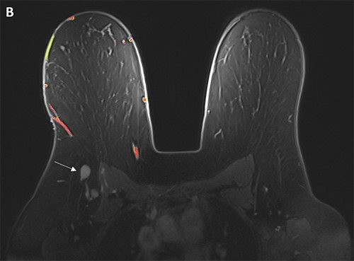
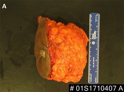
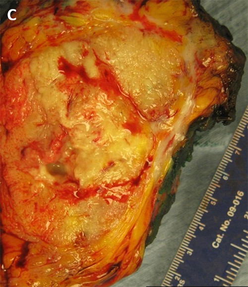
Figure 5. (A, B) Specimen with skin and associated abscess. (C) A crosssection of the specimen reveals a well-circumscribed lesion. Black ink denotes the margin.

Surgical pathology revealed a 9.5 cm tumor with negative margins, and one of two sentinel nodes had isolated tumor cells by immunohistochemistry. Based on the patient's stage and the current data suggesting that treatment for breast SCC be done in a manner similar to breast adenocarcinoma, our medical and radiation oncologists recommended adjuvant chemotherapy and adjuvant whole breast radiation.

In review of this case, a core needle biopsy of the patient's breast lesion prior to incision and drainage could have expedited the diagnosis had the radiologists been able to obtain tissue. Apart from this, the main obstacle that delayed her diagnosis was lack of insurance. Our breast center worked to connect the patient with resources so she could be seen as soon as they were aware of the patient, but the patient may have been diagnosed sooner had she been insured.

Discussion

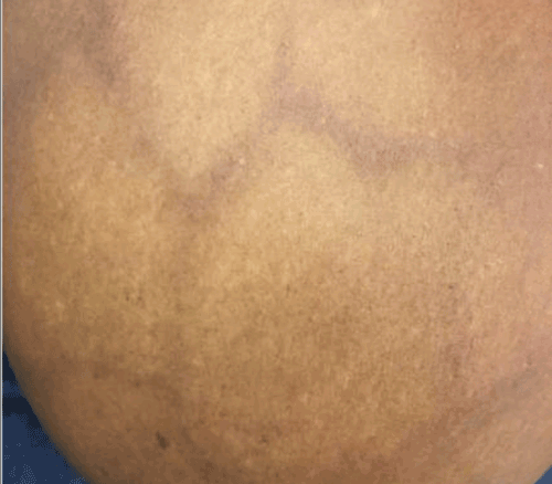
Breast abscesses commonly affect women of reproductive age and are often a complication of mastitis.4 These can be treated with antibiotics, aspiration, and/or incision and drainage depending on the abscess size and characteristics.4-7 Needle aspiration is usually considered first-line treatment for breast abscesses less than 5 cm, and multiple aspirations may be required. However, surgical incision and drainage can be considered for first-line therapy in large (>5 cm), multiloculated, or recurrent abscesses. Also, if an abscess recurs after multiple aspiration attempts, surgical drainage may be needed. As in our case, if there is no known underlying etiology for abscess recurrence, tissue sampling for pathology during surgical incision and drainage of chronic or complicated abscesses should be done to rule out underlying malignancy or benign inflammatory condition.

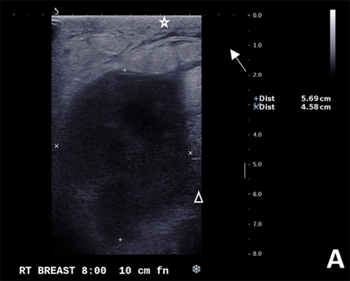
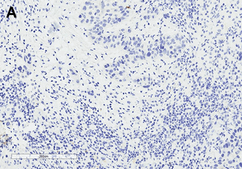
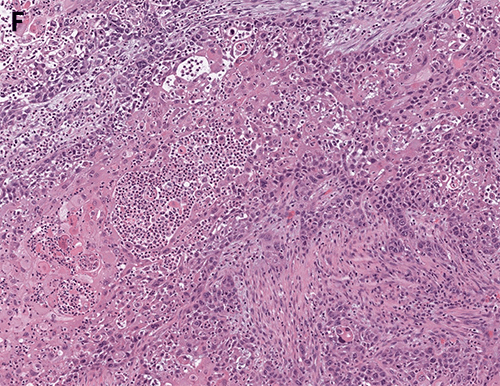
Although primary breast SCC is rare, based on the few reported cases, it is thought to be associated with chronic inflammation seen in benign breast diseases like abscesses, chronic cysts or sinuses, breast implants, or after radiation.3,8 It is hypothesized that SCC may arise from foci of squamous metaplasia within a preexisting adenocarcinoma or directly from mammary duct epithelium.2,5,8 To be diagnosed, primary SCC of the breast requires the tumor be free of other types of adenocarcinoma, not involve overlying skin, and not have another primary SCC.3,5,8 In general, primary breast SCCs are larger at time of presentation (>4 cm).1-3,5,8 Ultrasound may show a complicated cyst or inflammation, but otherwise primary breast SCCs do not have any known typical findings on mammogram.2,5,8 They are often hormone-receptor negative, as was the case in our patient, and some authors report that lymph node involvement is rare.1,5,8,9

Given the absence of specific management and treatment guidelines for primary breast SCC, management should be similar to that for breast adenocarcinoma.8-12 A surgical biopsy should be performed to establish the diagnosis, followed by clinical staging with imaging. Surgical intervention should be determined by clinical staging, often requiring partial mastectomy with sentinel lymph node biopsy to determine pathologic tumor staging. Adjuvant chemoradiation and hormone therapy should be used if indicated based on pathologic staging and receptor status.

Although prognosis of primary breast SCCs is controversial due to limited data, they are typically extremely aggressive tumors with courses comparable to poorly differentiated breast adenocarcinomas.1-3,5,8-12 Rates of locoregional relapse are high: one case series of 33 patients reported a median locoregional recurrence-free survival (RFS) of 20 months for patients without metastatic disease at diagnosis.8 In this study, 71 percent of these patients suffered relapse with median 14 month survival from the recognition of recurrent or metastatic disease. Their survival was somewhat worse than the still poor 64 percent overall five-year survival of patients in the SEER database. Due to the high rates of locoregional recurrence in this disease, early adjuvant radiation therapy is thought to be prudent despite reports of frequent recurrence in irradiated fields. Adjuvant chemotherapy is used regularly given the aggressive nature, but risk of distant metastasis remains high in SCC.8,9 Historically, anthracycline containing regimens have been the standard; however, the use of carboplatin and taxanes have biologic plausibility and have been employed.

Conclusion

Primary breast SCC is a rare and often aggressive cancer. In the absence of clear management guidelines for this tumor, medical and surgical management should follow guidelines for breast adenocarcinoma.

Lessons Learned

Given primary breast SCC can arise in the setting of benign chronic inflammatory conditions like breast abscess, it is important to maintain a high index of suspicion for an underlying inflammatory condition in patients who appear to have complicated cases of such benign diseases. The differential diagnosis for complicated breast abscess includes breast SCC as well as invasive carcinoma, granulomatous mastitis, and systematic lupus erythematosus. For this reason, any complex breasts lesions that requires surgical incision and drainage should be biopsied.

Authors

Bryce E. Haac, MDa; Rachel E. White, MDb; Nikki Tirada, MDc; Naomi Hardy, MDb; Yvonne Rasko, MDa; Elizabeth Nichols, MDd; Paula Rosenblatt, MDe; Sergio Dromi, MDc; Olga Ioffe, MDb; and Emily Bellavance, MDa

Correspondence Author

Dr. Bryce Haac
Department of Surgery
University of Maryland Medical Center
22 S. Greene St., 8th Floor
Baltimore, MD 212019
bhaac@som.umaryland.edu

Author Affiliations

  1. Department of Surgery
  2. Department of Anatomic Pathology
  3. Department of Diagnostic Radiology and Nuclear Medicine
  4. Department of Radiation Oncology
  5. Department of Hematology and Oncology

University of Maryland School of Medicine
Baltimore, MD

Disclosure Statement

The authors have no conflicts of interest to disclose.

References

  1. Gupta C, Malani AK, Weigand RT, Rangineni G. Pure primary squamous cell carcinoma of the breast: a rare presentation and clinicopathologic comparison with usual ductal carcinoma of the breast. Pathol Res Pract. 2006; 202(6):465-469.
  2. Flikweert ER, Hofstee M, Liem MSL. Squamous cell carcinoma of the breast: a case report. World J Surg Oncol. 2008; 6:135.
  3. Behranwala KA, Nasiri N, Abdullah N, Trott PA, Gui GPH. Squamous cell carcinoma of the breast: clinicopathologic implications and outcome. EJSO 2003;29:386-389.
  4. Kataria K, Srivastava A, Dhar A. Management of lactational mastitis and breast abscesses: review of current knowledge and practice. Indian J Surg. 2013 Dec;75(6):430-435.
  5. Seddik Y, Brahmi SA, Afqir S. Primary squamous cell carcinoma of the breast: a case report and review of literature. Pan Afr Med J. 2015;20:152.
  6. Lam E, Chan T, Wiseman SM. Breast abscess: evidence based management recommendations. Expert Rev Anti Infect Ther. 2014 Jul; 12(7): 753-762
  7. Barron AU, Luk S, Phelan HA, Williams BH. Do acute-care surgeons follow best practices for breast abscess management? A single-institution analysis of 325 consecutive cases. J Surg Res. 2017 Aug;216:169-171.
  8. Hennessy BT, Krishnamurthy S, Giordano S, Buchholz TA, Kau SW, Duan Z. Squamous cell carcinoma of the breast. J Clin Oncol. 2005 Nov 1;23(31):7827-7835.
  9. Behranwala KA, Nasiri N, Abdullah N, et al. Squamous cell carcinoma of the breast: Clinico-pathologic implications and outcome. Eur J Surg Oncol. 29:386,2003-389
  10. Cardoso F, Leal C, Meira A, et al: Squamous cell carcinoma of the breast. Breast. 9:315,2000-319
  11. Lafreniere R, Moskowitz LB, Ketcham AS: Pure squamous cell carcinoma of the breast. J Surg Oncol. 31:113,1986-119
  12. Badge SA, Gangane NM, Shivkumar VB, Sharma SM. Primary squamous cell carcinoma of the breast. Int J Appl Basic Med Res. 2014 Jan-Jun;4(1):53-55.