Unsupported Browser
The American College of Surgeons website is not compatible with Internet Explorer 11, IE 11. For the best experience please update your browser.
Menu
Become a member and receive career-enhancing benefits

Our top priority is providing value to members. Your Member Services team is here to ensure you maximize your ACS member benefits, participate in College activities, and engage with your ACS colleagues. It's all here.

Become a Member
Become a member and receive career-enhancing benefits

Our top priority is providing value to members. Your Member Services team is here to ensure you maximize your ACS member benefits, participate in College activities, and engage with your ACS colleagues. It's all here.

Membership Benefits
ACS
Case Reviews in Surgery

A Case of Retroperitoneal Schwannoma: Determination of Appropriate Timing for Surgical Resection

A Case of Retroperitoneal Schwannoma: Determination of Appropriate Timing for Surgical Resection

March 2, 2020

Abstract

Background

We present a case of a 61-year-old woman with a history of pelvic pain secondary to a chronic retroperitoneal mass that presented with a large, symptomatic recurrent ventral hernia. The patient underwent a concomitant elective resection of the retroperitoneal mass and was found to have a benign schwannoma on pathology.

Summary

Benign schwannomas are solitary, well-encapsulated, slow-growing masses rarely found in the retroperitoneum. Preoperative diagnosis is difficult, but can be made using core needle biopsy. Pathology and immunohistochemical analysis of the excised specimen are required for final diagnosis and show diffuse cytoplasm S-100 positivity.

Conclusion

Pre-operative diagnosis of retroperitoneal masses are challenging due to vague clinical presentation and lack of characteristic imaging findings. Although surgical excision is the definitive treatment of choice for schwannomas, conservative management with serial imaging is appropriate without risk factors for malignant transformation.

Keywords

Sarcoma, retroperitoneal mass, schwannoma


Case Description

We present a 61-year-old woman who was referred for evaluation due to an incidental finding of interval enlargement of a soft tissue retroperitoneal mass seen on computed tomography (CT) of the abdomen and pelvis. The patient had multiple medical comorbidities, including congestive heart failure, pacemaker placement secondary to sick sinus syndrome, chronic obstructive pulmonary disease, deep vein thrombosis, obesity, hypertension, and insulin-dependent diabetes. She also had an extensive past surgical history including antrectomy with partial gastrectomy, cholecystectomy, hysterectomy, and multiple prior open incisional hernia repairs with mesh. The patient had been diagnosed with this asymptomatic right pelvic soft tissue mass of uncertain etiology six years prior and had opted for conservative management with serial surveillance. The patient had two CT scans between 2012 and 2016, which had demonstrated relative stability in size. The mass was mildly enhancing with contrast, homogenous in appearance, and bilobed with clear margins. CT also demonstrated a lack of invasion into surrounding structures, necrosis, calcifications or septations. It was felt to be benign based on these findings as well as relative stability in size until 2017 when the mass increased ~1.5cm in the anterior-posterior and ~0.8cm in the craniocaudal dimensions. Thus, a slow growing malignant neoplasm could not be excluded, and the patient was subsequently referred for a CT-guided core-needle biopsy with interventional radiology.

Histopathology findings of core needle biopsy demonstrated spindle cells in fascicles with scattered mild chronic inflammatory infiltrates, suggesting a benign etiology. No glandular squamous differentiation was noted, although the spindle cell proliferation appeared to be slightly infiltrative into surrounding associated dense fibrovascular tissue. Immunoperoxidase staining was negative for desmin and CD34, weakly positive for pancytokeratin, and diffusely strongly positive for S-100. This was consistent with a schwannoma, however weakly positive staining for pancytokeratin only marginally excludes carcinoma. Given the patient's multiple comorbidities and lack of symptoms at that time, the decision was made not to pursue aggressive surgical management.

Unfortunately, the patient then developed progressively worsening abdominal distension and pelvic discomfort in 2018, and a repeat abdominal CT scan was obtained by the patient's primary care physician. The retroperitoneal mass remained stable in the year interim, however a slow growing neoplasm remained on the differential given the history of interval enlargement and weak pancytokeratin staining on histopathologic staining. Imaging findings were also remarkable for development of a large recurrent incisional hernia (Figure 1). Given the patient's pelvic pain presumed to be secondary to this persistent mass, in conjunction with her recurrent hernia, the patient was amenable to proceeding with abdominal wall hernia repair with concurrent resection of the right retroperitoneal mass.

Figure 1. Large right retroperitoneal mass (a) and associated large anterior wall abdominal hernia (b)

The patient was taken to the operating room for hernia repair with excision of mesh and resection of retroperitoneal mass under general anesthesia with endotracheal intubation. A longitudinal midline incision was made along the scar from her previous surgery. The hernia sac was identified and entered, revealing extensive small bowel adhesions which were carefully dissected, and extensive lysis of adhesions was performed from the ligament of Treitz to the ileocecal valve. The edges of the hernia were released, and the prior mesh was excised. The retroperitoneal mass was then carefully dissected from the right pelvis. Identification of the ureter was performed to prevent inadvertent injury. Dissection was complicated by the close proximity to the iliac vessels; however, a plane was easily created through mostly blunt dissection. The mass was able to be removed with capsule intact. The midline abdominal wall hernia was repaired with a large sublay composite mesh placement and the operation was concluded.

Figure 2. Retroperitoneal mass gross specimen

Gross examination of the excised specimen revealed a smooth appearing mass with focal adherent adipose tissue measuring 10.2 x 5.0 x 4.5cm (Figure 2). Cut surface revealed a central nodular lesion surrounded by compressed fibrous tissue. A nerve fiber was seen wrapping around the periphery of the tissue. Microscopic sectioning demonstrated bland spindle cells with wavy contours as well palisading nuclei and Verocay bodies (Figure 3). Focal areas of both dense and loose cellularity were seen throughout the lesion. Immunohistochemical staining was diffusely positive for S-100 protein.

Figure 3. Nerve fiber seen wrapping lesion peripherally (a) spindle cells with palisading nuclei (b) Antoni A with high cellularity (c) Antoni B with loose cellularity (d)

The patient had an uncomplicated postoperative course, with immediate improvement in her abdominal and pelvic pain. She was discharged home but then readmitted to the hospital two weeks later with CHF exacerbation and abdominal wall seroma, which was treated with percutaneous drainage. During her hospital stay she was also treated for mucosal and vaginal candidiasis. She tolerated these treatments well, improved clinically and was discharged home. She followed up in the office six months postoperatively and is doing well with complete resolution of her pelvic pain and no evidence of mass or hernia recurrence.

Discussion

Schwannomas are tumors of mesenchymal origin that arise from peripheral nerve sheath cells.1 These typically benign, solitary, and well-encapsulated neoplasms rarely arise from peripheral nerve fibers, with an estimated prevalence of 1-3%.1,3 Schwannomas more commonly manifest in the head and neck, extremities, mediastinum, and trunk.1 Although schwannomas can occur in all age groups, they are found predominantly in women in the 5th and 6th decades of life.2 Retroperitoneal tumors are rare, but are usually found incidentally. They frequently arise from the paravertebral region in the retroperitoneum, in relation to the nerve of origin, though neurologic symptoms are rare. Clinical presentation may be the result of compression on local structures, and the propensity for these tumors to present at late stages (with compressive symptoms) is a result of the compliance of the retroperitoneal space.1 In this patient's case, it was not clear which was the peripheral nerve of origin, however given its proximity it may have been branches of the femoral nerve, genitofemoral or obturator nerve. In contrast, patients may complain of nonspecific symptoms such as vague abdominal or pelvic pain.

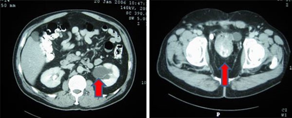
Imaging modalities that may assist with preoperative workup include CT and/or magnetic resonance imaging (MRI). CT scans usually reveal well-defined, heterogeneous lesions and may demonstrate cystic, necrotic, or hemorrhagic components frequently with internal calcification. MRI may be particularly useful in identifying vascular architecture suggestive of malignancy, although this is nonspecific.1 Although some institutions use adjunct final needle aspiration cytology to aid in diagnosis, the varying characteristics of these lesions invariably lower the contribution of the results and such an approach should be limited to centers that have developed specific expertise in these protocols. As a result, core needle biopsies should be obtained as part of the standard diagnostic workup as per the European Society for Medical Oncology guidelines.

Malignant transformation to neurofibrosarcoma is rarely seen, however has been reported to occur following surgical resection of histologically benign schwannomas. The development of malignant changes is more likely to occur in patients with Von Recklinghausen disease.4 Histologic features suggestive of malignant potential include high mitotic count, pleomorphism, and neovascularization.2 Postoperative monitoring with serial surveillance using annual CT scans is recommended to monitor carefully for recurrence or malignancy.1

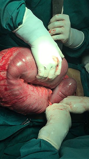
Complete surgical excision with negative margins is the recommended curative treatment, as postoperative pathology and immunohistochemistry is needed for final diagnosis. In addition, schwannoma responds poorly to radiation and chemotherapy and so adjuvant therapies are not an option. An en bloc excision of adjacent structures may be warranted but was not necessary in this case. The patient's hernia was repaired with synthetic mesh, given the need for tension free repair in the setting of recurrent hernia, and the lack of contamination. Gross examination of schwannomas usually reveal solitary, well-circumscribed, smooth tumors. Histologic features include a biphasic pattern with areas of high (Antoni type A) and loose cellularity (Antoni type B).2,3,4 Nuclear palisading and Verocay bodies are classically seen, although their presence may be variable.2 Tumors show diffuse cytoplasm positivity for S-100 protein.

Conclusion

Our patient had multiple comorbidities that precluded aggressive surgical management of an initially asymptomatic retroperitoneal mass. Interval surveillance allowed for appropriate recognition of mass enlargement, as well as symptoms related to the mass. The development of a large, recurrent, symptomatic incisional hernia necessitated intervention, allowing for concurrent resection of a known retroperitoneal mass.

Lessons Learned

The purpose of serial surveillance is ultimately to determine appropriateness and timing of intervention. Pre-operative diagnosis of retroperitoneal schwannomas is challenging given non-specific radiographic features and low yield of percutaneous biopsies. Definitive management is with surgical excision, which allows for both treatment, and diagnosis with postoperative pathology and immunohistochemistry.

Authors

Monica K Zipple, MD
Department of General Surgery
St Joseph Mercy Oakland
Pontiac, MI

Anna L Shu, DO
Department of General Surgery
St Joseph Mercy Oakland
Pontiac, MI

Sujeivan Mahendram, MS
Department of General Surgery
St Joseph Mercy Oakland
Pontiac, MI

Malik E McKany, MD
Department of General Surgery
St Joseph Mercy Oakland
Pontiac, MI

Correspondence

Monica Zipple, MD
44405 Woodward Avenue
Pontiac, MI 48341
(989) 506-1533
mzipple@gmail.com

Disclosure of Conflicts

The authors have no conflicts of interest to disclose.

References

  1. Holbrook C, Saleem N. Retroperitoneal schwannoma: an unusual cause of abdominal distention. BMJ Case Reports 2017; 2017: bcr-2017-220221.
  2. Narasimha A, Kumar H, Kalyani R, Madan M. Retroperitoneal cystic schwannoma: A case report with review of literature. J Cytol 2010; 27(4): 136-139.
  3. Kapan M, Önder A, Gümüş M, Gümüş H, Girgin S. Retroperitoneal schwannoma. J Surg Case Rep 2011;10: 1–1.
  4. Goh B, Tan Y, Chung Y, Chow P, Ooi L, Wong W. Retroperitoneal schwannoma. Am J Surg 2006; 192(1): 14–18.