November 23, 2020
Lipomas are benign tumors of mature fat cells that most commonly affect areas with abundant adipose tissue. When they occur in the hand, they most commonly affect the hypothenar and thenar areas, with less than one percent occurring on fingers.
We report a case of a large lipoma of the middle finger involving the volar aspect of the proximal finger. The mass had been present for five years prior to presentation, and the patient complained of tenderness without associated paresthesia. An MRI was consistent with the diagnosis of lipoma. During excision, it was noted that the mass had grown between the digital neurovascular bundle separating the digital nerve from the proper digital artery. The artery remained dorsal to the neoplastic growth, while the nerve had been forced toward the volar portion of the finger. Pathology confirmed the diagnosis of lipoma, and the patient did well following the procedure with no deficits in range of motion or sensation.
Large digital lipomas, while rare, have the potential to separate the neurovascular architecture of the finger. They should be included in the differential diagnosis of large digital tumors.
Lipoma; tumor; finger; digital nerve; paresthesia; digital artery; hand
Lipomas are benign tumors of mature fat cells that most commonly affect areas with abundant adipose tissue. When they occur in the hand, they most commonly affect the hypothenar and thenar areas with less than one percent occurring on fingers.1,2 We report a case of a large 3.5 x 2.5 cm lipoma of the middle finger involving volar aspect of the proximal middle finger. Intraoperative dissection revealed the tumor growth had separated the proper digital artery and nerve.
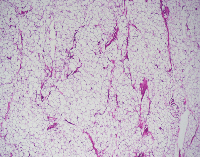
A 61-year-old, right-hand-dominant female was seen in the clinic for a five-year history of a left middle finger mass. The patient had complaints of mild tenderness directly over the mass, no distal paresthesia, and full range of motion with mild mass impingement at full flexion. Examination revealed a soft, well-circumscribed mobile mass involving the volar aspect of the middle finger with normal appearing skin overlying it (Figure 1A). The mass was confined to the proximal phalanx but did extend dorsally along the radial and ulnar side of the finger. Magnetic resonance imaging (MRI) was consistent with lipoma, revealing a homogenous, multi-lobular encapsulated mass 2.5 by 3.5 cm in size (Figure 1B). The patient was taken to the OR for excision through a Bruner incision. Notably, the mass had grown between the digital neurovascular bundle, separating the digital nerve from proper digital artery (Figure 1C and Figure 1D). The artery remained dorsal to the neoplastic growth, while the nerve had been forced toward the volar portion of the finger. Pathology of the specimen revealed numerous mature adipocytes, confirming the rare diagnosis of digital lipoma (Figure 2A). Postoperatively, the patient did well and healed completely. She achieved painless, full range of motion along with normal sensibility.
Figure 1. A) Gross presentation of the digital lipoma. B) T1-MRI image of the mass showing homogenous, multi-lobular encapsulated mass 2.5 by 3.5cm in size. C and D) Intraoperative images showing the nerve on the volar aspect of the mass and artery lying dorsally.
Figure 2. Pathology section of digital lipoma. Mature adipocytes are present throughout the visual field with uniform nuclei confirming the diagnosis of digital lipoma.
Lipomas are the most common benign soft-tissue neoplasm in adults.3 They are most often found in the trunk or upper extremity, and they most often present in subcutaneous planes of areas with increased adiposity. They rarely involve the hand, and masses involving the fingers may represent less than one percent of all lipomata.1 Only a handful of digital lipomas are described in the literature, but greater than one-third (35.14 percent) involved the middle finger.2,4 Reports associate these tumors with neuropathic pain, paresthesia, restricted range of motion, bony erosion, and decreased nail bed perfusion.4 Histologically, they do not differ from lipomas in more classical anatomical locations and consist of mature adipocytes covered by a thin fibrous capsule. Gross examination of lipomas, most typically show well-circumscribed masses with a yellow appearance.
Given their rarity, lipomas are often excluded from the differential diagnosis of slow-growing, painless masses of the finger, which are more commonly attributed to mucoid cysts, inclusion cysts, or giant cell tumors.5 In fact, recent metanalyses of these lesions revealed that most were not diagnosed or considered until after excision.4 One case reported involvement of the digital nerve6 but failed to describe its relationship to the artery. Several others have associated the neoplasms with middle finger paresthesia and pain,4 which may be attributable to involvement or entrapment of the neurovascular bundle.
This current case is notable in respect to the size of the lipoma and separation of the digital artery and nerve, a phenomenon not previously described in the literature (to the best of the authors’ knowledge). With regard to size, only two previously described cases involving the middle finger were larger than the mass found in our case.7,8 This previously unreported involvement of the neurovascular structures should be considered during the excision of large lipomas and may be a source for commonly reported paresthesia in digital lipomas.
Lipomas should be included in the differential of large digital tumors, especially when associated imaging appears homogenous. Digital lipomas, while rare, have the potential to separate the neurovascular architecture of the finger. This involvement may explain previously described reports of distal paresthesia occurring with these tumors and should be considered during excision.
Digital lipomas have the potential to separate the neurovascular architecture of the finger and may be associated with distal paresthesia reported with these rare tumors.
McCulloch ILa; McClellan WTb
Ian L. McCulloch, MD, MRes
16 Willow Lane
Morgantown, WV 26505
Phone: (304) 237-0173
E-mail: imccullo@mix.wvu.edu
a West Virginia University, School of Medicine, Morgantown, WV 26506
b McClellan Plastic Surgery, Morgantown, WV 26508
The authors have no conflicts of interest to disclose.