Discussion
Amyand hernia, or the appendix incarcerated within an inguinal hernia, was first described in 1735 by Claudius Amyand.1-3 There are also case reports of appendix incarceration within incisional hernias,4-7 femoral hernias (De Garengeot hernia),8,9 and within a right lower quadrant port site.4,10 The incidence of appendiceal hernia is an uncommon occurrence, and the development of complications, including inflammation, perforation, abscess, or fistula formation is even more rare (<0.1 percent).5,11 Due to the variety of locations and range of complications that have been described in appendiceal hernias, the patient presentation may differ significantly from classic appendicitis. Additionally, presenting signs and symptoms may vary significantly between affected patients. Thus, a high index of suspicion is required for appropriate diagnosis and timely surgical management.
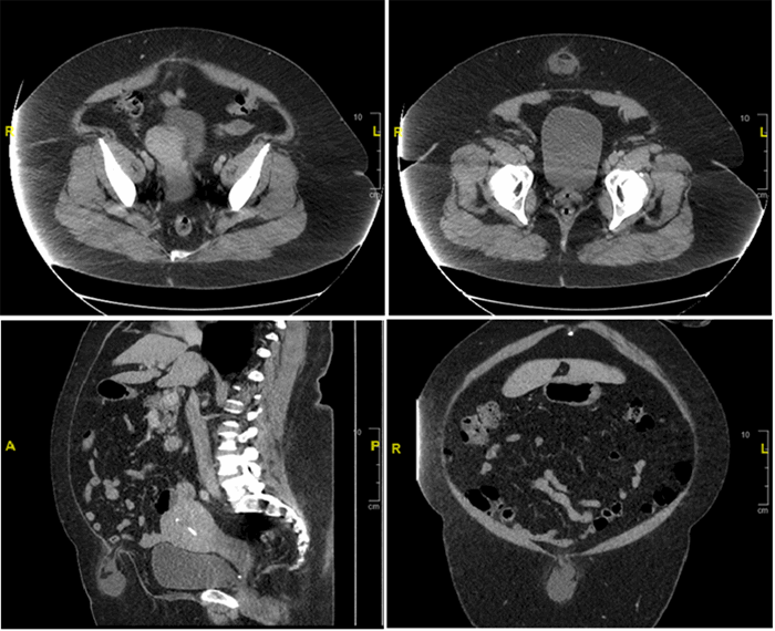
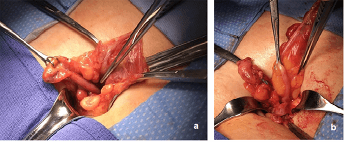
This paper represents the first report of an appendix incarcerated within a midline incisional port site hernia, treated prior to the development of complications. The risk of developing a port-site hernia after cholecystectomy range from 0.3 to 5.4 percent.12-14 Several risk factors have been associated with the development of a PSH, including an increased body mass index, trocar diameter and design, older age, and longer duration of surgery.12 Location of the port is also a contributory factor, with the most common site being at the umbilicus or midline, due to the weakness of the linea alba.15 While this patient was obese, further evaluation of other risk factors related to her initial surgery was inconclusive given that her cholecystectomy had been performed at an outside institution. Other pertinent contributing factors include the proximity of the cecum and appendix to the fascial defect (Figure 1) and length of the appendix. While the pathology measurement of the appendix was 7 cm in length, it can be seen to stretch to a much longer length in Figure 2. Port site hernia should remain on the surgeons’ radar for potentially avoidable complication following laparoscopic surgery and can be avoided with appropriate closure of fascial defects and risk factor modification.
Concomitant primary repair of the PSH was possible in this case, given the lack of field contamination and the small size of the hernia defect (<1 cm). Considering the patient’s risk factors for recurrence, if the fascial defect had been larger, then repair with ultra-lightweight mesh reinforcement may have been considered. However, we should note there is conflicting literature regarding the use of mesh repair in repair of clean-contaminated hernias.16,17 If there had been any concern for contamination of the wound, then the authors would have considered leaving the skin open to heal by secondary intention or loose closure of the wound with staples.
A similar patient presenting with an incarcerated incisional PSH thought to contain simply preperitoneal or omental fat may have been discharged with plans for elective hernia repair. This type of delay in management of an appendiceal hernia may ultimately result in a more advanced pathology, including ischemia, perforation, abscess or fistula formation, for which the surgical management may involve a significantly more morbid procedure. This scenario would need to be approached similarly to emergent management of strangulated bowel, and definitive management of the hernia would be left to the discretion of the operating surgeon. This patient was able to undergo a definitive surgical operation with a short hospital stay and no adverse events.
Conclusion
The appendix can become incarcerated with a variety of different hernias and locations and may be difficult to diagnose preoperatively. This represents the first report of an appendix incarcerated within a midline port site treated prior to the development of complications. Early recognition of appendiceal hernia allows for management with appendectomy and herniorrhaphy with minimal morbidity to the patient.
Lessons Learned
An incarcerated hernia containing the appendix is a rare type of hernia, and the presentation can differ significantly from classic appendicitis that may increase incidence of late presentation and delay in diagnosis or treatment. Preoperative diagnosis of this rare type of hernia may be missed by physical exam and imaging like abdominal CT scan. PSH is also extremely rare and should be a remain high on the surgeon’s radar of potential complications after laparoscopic surgery. Surgical management should be the preferred treatment of incarcerated appendiceal hernia and should be performed as soon as diagnosis is suspected in order to prevent development of significant complications.
Authors
Zipple MK; Chonghasawat A; Caugh D; Yaldo B
Author Affiliation
St. Joseph Mercy Oakland
Department of General Surgery
Pontiac, MI 48341
Corresponding Author
Monica Zipple, MD
44405 Woodward Avenue
Pontiac, MI 48341
Phone: (989) 506-1533
Email: mzipple@gmail.com
Disclosure Statement
The authors have no conflicts of interest to disclose.
References
- Amyand C. Of an inguinal rupture, with a pin in the appendix caeci, incrusted with stone, and some observations on wounds in the guts. Philos Trans R Soc Lond. 1736; 39: 329-336.
- Shaban Y, Elkbuli A, McKenney M, Boneva D. Amyand's hernia: A case report and review of the literature. Int J Surg Case Rep. 2018;47:92-96. doi:10.1016/j.ijscr.2018.04.034
- Ivanschuk G, Cesmebasi A, Sorenson EP, Blaak C, Loukas M, Tubbs SR. Amyand's hernia: a review. Med Sci Monit. 2014;20:140-146. Published 2014 Jan 28. doi:10.12659/MSM.889873
- Sugrue C, Hogan A, Robertson I, Mahmood A, Khan WH, Barry K. Incisional hernia appendicitis: A report of two unique cases and literature review. Int J Surg Case Rep. 2013;4(3):256-258. doi:10.1016/j.ijscr.2012.12.006
- Kane ED, Bittner KR, Bennett M, Romanelli JR, Seymour NE, Wu JJ. A case report of unexpected pathology within an incarcerated ventral hernia. Int J Surg Case Rep. 2017;38:61-65. doi:10.1016/j.ijscr.2017.07.004
- Kler A, Hossain N, Singh S, Scarpinata R. Vermiform appendix within incisional hernia. BMJ Case Rep. 2017;2017:bcr2017221216. Published 2017 Aug 20. doi:10.1136/bcr-2017-221216
- Galiñanes EL, Ramaswamy A. Appendicitis found in an incisional hernia. J Surg Case Rep. 2012;2012(8):3. Published 2012 Aug 1. doi:10.1093/jscr/2012.8.3
- Konofaos P, Spartalis E, Smirnis A, Kontzoglou K, Kouraklis G. De Garengeot's hernia in a 60-year-old woman: a case report. J Med Case Rep. 2011;5:258. Published 2011 Jun 30. doi:10.1186/1752-1947-5-258
- Bidarmaghz B, Tee CL. A case of De Garengeot hernia and literature review. BMJ Case Rep. 2017;2017:bcr2017220926. Published 2017 Sep 7. doi:10.1136/bcr-2017-220926
- Menenakos Ch, Tsilimparis N, Guenther N, Braumann C. Strangulated appendix within a trocar site incisional hernia following laparoscopic low anterior rectal resection. A case report. Acta Chir Belg. 2009;109(3):411-413. doi:10.1080/00015458.2009.11680450
- Michalinos A, Moris D, Vernadakis S. Amyand's hernia: a review. Am J Surg. 2014;207(6):989-995. doi:10.1016/j.amjsurg.2013.07.043
- Bunting DM. Port-site hernia following laparoscopic cholecystectomy. JSLS. 2010;14(4):490-497. doi:10.4293/108680810X12924466007728
- Lee JH, Kim W. Strangulated small bowel hernia through the port site: a case report. World J Gastroenterol. 2008;14(44):6881-6883. doi:10.3748/wjg.14.6881
- Tonouchi H, Ohmori Y, Kobayashi M, Kusunoki M. Trocar site hernia. Arch Surg. 2004;139(11):1248-1256. doi:10.1001/archsurg.139.11.1248
- Pamela D, Roberto C, Francesco LM, et al. Trocar site hernia after laparoscopic colectomy: a case report and literature review. ISRN Surg. 2011;2011:725601. doi:10.5402/2011/725601
- Choi JJ, Palaniappa NC, Dallas KB, Rudich TB, Colon MJ, Divino CM. Use of mesh during ventral hernia repair in clean-contaminated and contaminated cases: outcomes of 33,832 cases. Ann Surg. 2012;255(1):176-180. doi:10.1097/SLA.0b013e31822518e6
- Fischer JP, Basta MN, Krishnan NM, Wink JD, Kovach SJ. A Cost-Utility Assessment of Mesh Selection in Clean-Contaminated Ventral Hernia Repair. Plast Reconstr Surg. 2016;137(2):647-659. doi:10.1097/01.prs.0000475775.44891.56


