Unsupported Browser
The American College of Surgeons website is not compatible with Internet Explorer 11, IE 11. For the best experience please update your browser.
Menu
Become a member and receive career-enhancing benefits

Our top priority is providing value to members. Your Member Services team is here to ensure you maximize your ACS member benefits, participate in College activities, and engage with your ACS colleagues. It's all here.

Become a Member
Become a member and receive career-enhancing benefits

Our top priority is providing value to members. Your Member Services team is here to ensure you maximize your ACS member benefits, participate in College activities, and engage with your ACS colleagues. It's all here.

Membership Benefits
ACS
Case Reviews in Surgery

Complex Abdominal Wound Closure Using Modified DermaClose System in Critically Ill Cardiac Patient with Open Abdomen

Damage control laparotomy is a commonly used strategy when dealing with intra-abdominal catastrophe.

July 22, 2021

Abstract

Background

Damage control laparotomy is a strategy used when dealing with intra-abdominal catastrophe. The open abdomen, one of the elements of this strategy, decreases the incidence of abdominal compartment syndrome and allows for serial second-look laparotomies to stabilize the intra-abdominal process prior to definitive abdominal wall closure. While direct closure of fascia is ideal, this is sometimes not possible, and other methods of definitive abdominal wall closure must instead be explored.

Summary

This case report details the clinical presentation and treatment of a 52-year-old male with mixed ischemic and non-ischemic cardiomyopathy with an ejection fraction of 10 percent and dual-chamber implantable cardioverter-defibrillator (ICD) who deteriorated into ventricular tachycardia (VT) storm and biventricular cardiogenic shock with multiple organ system failure requiring VA-ECMO (veno-arterial extracorporeal membrane oxygenation). He was transitioned to bilateral CentriMag® devices but developed a large, right retroperitoneal hematoma requiring decompressive laparotomy. Direct fascial closure was not possible, and thus his abdomen was closed with an inlay bridge biologic mesh, and a wound vacuum was placed over the mesh. Due to the large skin and subcutaneous tissue gap between skin flaps not amenable to primary closure, two DermaClose® systems were put into the skin in combination with wound vacuum therapy. The DermaClose® was originally designed for the closure of small extremity wounds. The DermaClose® system allowed for abdominal wall skin flap primary closure.

Conclusion

This is the first case report detailing our novel modification of the DermaClose® system in combination with wound vacuum therapy on the abdominal wall in a critically ill cardiac patient with an open abdomen and a large skin flap gap. With our modification, our patient was able to undergo successful abdominal wall closure and heart transplantation.

Key Words

abdominal compartment syndrome; open abdomen


Case Description

Damage control laparotomy is a commonly used strategy when dealing with intra-abdominal catastrophe. The open abdomen is one of the elements of this strategy. The open abdomen with temporary abdominal wall closure is indicated in the presence of the "triad of death" (coagulopathy, acidosis, and hypothermia) and situations associated with an increased risk of postoperative abdominal compartment syndrome (ACS). In conditions such as these, immediate closure of the abdomen may further compromise the patient's physiology.

Abdominal compartment syndrome is defined as an increase in intra-abdominal pressure leading to decreased cardiac preload, and increased intra-thoracic pressure resulting in cardiovascular collapse, acute respiratory failure, and acute kidney injury. The causes of ACS are multiple and include intra-abdominal trauma, ruptured abdominal aortic aneurysm, retroperitoneal hemorrhage, acute pancreatitis, burns, excessive fluid resuscitation, and sepsis, among others.1 Once the abdomen has been opened, various methods have been described to protect the abdominal contents and preserve the abdominal domain. These methods include different modifications of temporary abdominal wall dressings with negative pressure wound therapy (wound vacuum), Wittmann patch with transabdominal wall traction (TAWT), and other systems to preserve the abdominal domain.2,3

Definitive abdominal wall closure should not be delayed, however, as early closure of the abdomen has been shown to improve survival in these patients significantly.4,5 Once the patient's intra-abdominal conditions requiring an open abdomen are controlled and overall physiology normalized, final abdominal closure should be attempted.5 Direct closure of the fascia and skin is ideal. Still, this is not feasible in many cases due to loss of abdominal domain, extensive peritoneal adhesions, and excessive edema of intraabdominal organs and retroperitoneal structures. Therefore, other permanent abdominal wall closure techniques may be used alone or in combination: skin grafting over the granulated open abdomen, mesh-augmented closure with either synthetic or biologic products as an inlay or underlay, and component separations.68 Of note, pedicle or free flap abdominal wall reconstruction should be mentioned as a last resort in the reconstructive ladder that should only be undertaken as part of a multidisciplinary team with plastic surgery involvement.8

This case report describes a novel method of definitive abdominal wall closure in a patient with both a right ventricular assist device (RVAD) and left ventricular assist device (LVAD) who developed ACS secondary to a retroperitoneal hematoma necessitating decompressive laparotomy.

The patient is a 52-year-old male with a past medical history of mixed ischemic and non-ischemic cardiomyopathy with an ejection fraction of 10 percent and dual-chamber implantable cardioverter-defibrillator (ICD). He was transferred to our institution for evaluation of advanced cardiac support therapies (ventricular assist devices and orthotopic heart transplant) after deteriorating into ventricular tachycardia (VT) storm. Aggressive medical therapy for VT was unsuccessful, and he continued to deteriorate with VT storm and associated biventricular cardiogenic shock with multiple organ system failure (respiratory failure, shock liver, and hepatic encephalopathy) requiring peripheral VA-ECMO (veno-arterial extrcorporeal membrane oxygenation). This was achieved through 25 French right femoral vein cannula, 19 French right femoral arterial cannula, and 7 French antegrade catheter in superficial femoral artery for perfusion of the right lower extremity. He was transitioned eight days later to central cannulation with bilateral CentriMag® devices, and peripheral VA-ECMO cannulae were removed. Ten days after removal of ECMO cannulae, the patient developed an acute drop in his hematocrit resulting in an emergent computed tomography (CT) of his abdomen and pelvis. This showed a large right retroperitoneal hematoma without associated arterial pseudoaneurysm but presumed secondary to ECMO cannulation and systemic anticoagulation management. The patient developed the clinical picture of abdominal compartment syndrome and was emergently taken to the operating room for decompressive laparotomy with temporary abdominal wall closure. During exploration, the retroperitoneal hematoma was visualized but not evacuated as it was stable in size during the operation.

After the surgery, the patient was taken back to the cardiothoracic ICU for resuscitation. Forty-eight hours later, after extensive diuresis, the patient was taken to the operative room. The definitive abdominal fascia closure was impossible, given his tenuous cardiac function and excessive third spacing. Instead, we elected to place a Wittman patch with a transabdominal wall traction system and negative pressure dressing with plans for every other day tightening of the Wittmann patch. This is our standard approach to managing the difficult open abdomen. During the next 18 days, he returned to the operating room for abdominal washouts and Wittmann patch tightening. During this time, there were at least two episodes when the abdominal fascia was almost completely approximated. Unfortunately, the patient's hemodynamic status was so tenuous that every attempt to close the abdominal wall resulted in the clinical picture of abdominal compartment syndrome with RVAD suction events and hypotension. Each of these times, the Wittmann patch was loosened, and after optimization of the hemodynamic parameters, another Wittmann patch tightening was attempted without success. We hoped to achieve primary fascial closure, as otherwise, the patient could not be listed for a heart transplant until his abdomen was closed and healed.

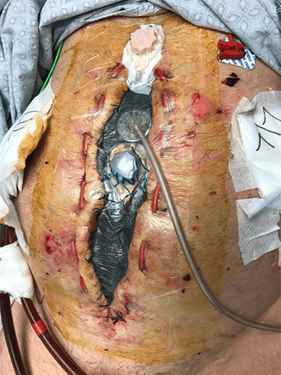
The decision was made to close the abdominal fascia with a biologic mesh and a 20 cm x 40 cm Strattice mesh that was placed in an inlay bridge fashion and sutured to the abdominal wall fascia given the patient's persistently tenuous hemodynamics (Figure 1). The size of the abdominal wall defect (20 cm x30 cm) did not allow us to close the skin primarily even after the creation of bilateral skin flaps. The complicating factor here was the presence of bilateral ECMO cannulas going through the mid-abdomen closely located to the midline laparotomy incision. A negative pressure vacuum dressing was applied on top of the Strattice mesh to cover the skin defect. We assumed that wound vacuum dressing would facilitate the growth of granulation tissue from the skin flaps over the mesh.

Figure 1. Abdominal closure with 20 cm x40 cm inlay Strattice mesh sutured to posterior rectus fascia

A few days later, due to lack of granulation of the subcutaneous tissue, two DermaClose® systems were put into the skin flaps with a wound vacuum dressing underneath. The skin anchors supplied with the DermaClose® system were not sufficient to be used as directed by the company recommendations as this tore through the skin due to the amount of tension. These skin anchors were replaced with 2-0 ETHIBOND® U-sutures placed through the skin flaps with the DermaClose® system intertwined in a shoelace-like manner (Figure 2). For the following week, the patient's DermaClose® was tightened at the bedside daily with anchoring 2-0 ETHIBOND® vertical mattress sutures placed in between to begin closing the wound. Over the week, the dimensions of the wound decreased from 20 cm x10 cm to 20 cm x4 cm (Figure 3). At this point, we changed to every other day tightening, and the wound was able to be closed entirely on day 11 after DermaClose® placement. Afterward, the patient had incisional wound vacuum changes every other day and was able to start working with physical therapy ten days later.

Figure 2. DermaClose® systems in place at superior and inferior poles of wound using anchoring 2-0 ETHIBOND® sutures in lieu of DermaClose® hooks

Figure 3. DermaClose® systems with wound vacuum in place overlying abdominal wound

Over the next few weeks, the wound slightly separated, and a wound vacuum dressing was applied. During vacuum changes, it was noted that the skin and soft tissue flaps were adhering to the underlying biologic mesh and the initiation of healthy granulation tissue. Approximately two months later, the patient's wound bed had sufficient granulation tissue (Figure 4), and he was taken for split-thickness skin graft (15 cm x 2 cm) with the left thigh as a donor site. Over time, the patient was able to stand and walk with PT. He underwent a successful heart transplant 17 days later.

Figure 4. Abdominal wound before application of split-thickness skin graft

Discussion

To our knowledge, this is the first case report publication detailing our modification of the DermaClose® system with wound vacuum therapy on the abdominal wall with an open abdomen. The DermaClose® system is a continuous external tissue expander that promotes rapid tissue expansion through continuous tension to approximate wounds. The DermaClose® system was traditionally designed for smaller wounds in the extremities, such as soft tissue wounds and fasciotomies, where closure is typically achieved in three to five days with decreased use of skin grafting.9 However, due to the patient's body habitus and poor cardiac status, creative solutions, namely the DermaClose® system, had to be explored to facilitate abdominal closure. Of note, it should be mentioned that we initially attempted primary fascial closure for 18 days to accelerate the patient's relisting for heart transplant with the understanding that the risk of enteroatmospheric fistula increases significantly after 7–10 days.2

We used large ETHIBOND® sutures to help pull the skin with subcutaneous tissue together combined with the DermaClose® system to cause tissue expansion. Eventually, due to the amount of tension required, we substituted the traditional DermaClose® hooks with ETHIBOND® suture loops to pull the skin edges more tightly. With aggressive diuresis, daily tightening, and negative pressure therapy, we were able to get a very large wound defect closed in a relatively short amount of time. With the patient’s abdominal wall closed, he could be listed again for a heart transplant, and he was successfully transplanted. He is now living independently at home following his heart transplant. He is awaiting further abdominal wall reconstruction as bridging with biologic mesh is known to result in a ventral hernia but is a viable option.10 It should be noted that thought was given to performing component separation as well as rotational flaps in consultation with plastic surgery. Still, the presence of bilateral ECMO cannulae made this type of reconstruction prohibitive.

Conclusion

Although time and resource-intensive, by using the DermaClose® system in a unique and modified way in combination with wound vacuum therapy, we were able to close this complicated patient's abdomen. While he did have small wound dehiscence requiring further therapy and ultimately a skin graft, closing the majority of his wound and only having a small wound vacuum meant that he could participate in physical therapy to be relisted for a heart transplant and ultimately transplanted. Without this system, we presume it would have taken a much longer period of time to granulate the wound, and time to listing would have been significantly delayed. Therefore, in more complicated patients, after traditional wound closure methods have been exhausted, this method is a viable alternative.

Lessons Learned

When traditional methods of abdominal wall closure in patients with open abdomen are contraindicated or have been exhausted, a novel method of abdominal wall closure using the DermaClose® system is a viable alternative.

Authors

Velez Ma; Bloom JAa; Reich JAb; Bugaev Na

Author Affiliations

  1. Tufts Medical Center, Department of Surgery, Boston, MA 02176
  2. Tufts Medical Center, Department of Anesthesiology, Boston, MA 02176

Corresponding Author

Joshua A. Bloom, MD
Tufts Medical Center Boston, Department of Surgery
800 Washington Street
Boston, MA 02176
Phone: (561) 445-4690
E-mail: jbloom1@tuftsmedicalcenter.org

Disclosure Statement

The authors have no conflicts of interest to disclose.

References

  1. Kääriäinen M, Kuokkanen H. Primary closure of the abdominal wall after "open abdomen" situation. Scand J Surg. 2013;102(1):20-24. doi:10.1177/145749691310200105
  2. Leppäniemi A, Tukiainen E. Reconstruction of complex abdominal wall defects. Scand J Surg. 2013;102(1):14-19. doi:10.1177/145749691310200104
  3. Demetriades D, Salim A. Management of the open abdomen. Surg Clin North Am. 2014;94(1):131-153. doi:10.1016/j.suc.2013.10.010
  4. Sharrock AE, Barker T, Yuen HM, Rickard R, Tai N. Management and closure of the open abdomen after damage control laparotomy for trauma. A systematic review and meta-analysis. Injury. 2016;47(2):296-306. doi:10.1016/j.injury.2015.09.008
  5. Cristaudo AT, Jennings SB, Hitos K, Gunnarsson R, DeCosta A. Treatments and other prognostic factors in the management of the open abdomen: A systematic review. J Trauma Acute Care Surg. 2017;82(2):407-418. doi:10.1097/TA.0000000000001314
  6. Kääriäinen M, Kuuskeri M, Helminen M, Kuokkanen H. Greater success of primary fascial closure of the open abdomen: a retrospective study analyzing applied surgical techniques, success of fascial closure, and variables affecting the results. Scand J Surg. 2017;106(2):145-151. doi:10.1177/1457496916665542
  7. Hultman CS, Pratt B, Cairns BA, et al. Multidisciplinary approach to abdominal wall reconstruction after decompressive laparotomy for abdominal compartment syndrome. Ann Plast Surg. 2005;54(3):269-275.
  8. Boukovalas S, Sisk G, Selber JC. Abdominal wall reconstruction: an integrated approach [published correction appears in Semin Plast Surg. 2018 Nov;32(4):199-202]. Semin Plast Surg. 2018;32(3):107-119. doi:10.1055/s-0038-1667062
  9. Potter BK, Freedman BA, Shuler MS. Fasciotomy wound management and closure. Techniques in Orthopaedics. 2012;27(1):62-66. doi:10.1097/bto.0b013e31824885fa
  10. Coccolini F, Roberts D, Ansaloni L, et al. The open abdomen in trauma and non-trauma patients: WSES guidelines. World J Emerg Surg. 2018;13:7. Published 2018 Feb 2. doi:10.1186/s13017-018-0167-4