Unsupported Browser
The American College of Surgeons website is not compatible with Internet Explorer 11, IE 11. For the best experience please update your browser.
Menu
Become a member and receive career-enhancing benefits

Our top priority is providing value to members. Your Member Services team is here to ensure you maximize your ACS member benefits, participate in College activities, and engage with your ACS colleagues. It's all here.

Become a Member
Become a member and receive career-enhancing benefits

Our top priority is providing value to members. Your Member Services team is here to ensure you maximize your ACS member benefits, participate in College activities, and engage with your ACS colleagues. It's all here.

Membership Benefits
ACS
Case Reviews in Surgery

Benign Schwannoma of the Adrenal Gland and Mediastinum: Case Report and Literature Review

January 11, 2022

Abstract

Background

A 61-year-old woman presented with an asymptomatic, incidental left adrenal mass and simultaneous mediastinal lymphadenopathy. A biochemical evaluation was negative for functional tumors; however, size and radiological characteristics were concerning for malignancy.

Summary

In the era of the widespread use of high-resolution computed tomography (CT) and magnetic resonance imaging (MRI), adrenal incidentalomas are increasingly diagnosed in clinical practice. In this paper, we present a case of a schwannoma, or a benign peripheral nerve sheath tumor, manifesting as an adrenal incidentaloma and synchronous mediastinal mass. This patient was managed with a core needle biopsy of the mediastinal mass and subsequent operative adrenalectomy—the final pathology of both demonstrated benign peripheral nerve sheath tumor or schwannoma.

Conclusion

Peripheral nerve sheath tumors are a well-recognized, albeit rare, category of adrenal incidentalomas. Although they have very low malignant potential, these cannot be easily distinguished preoperatively. Therefore, operative resection is still ideal in ambiguous cases due to the inability to rule out adrenocortical carcinoma preoperatively.

Key Words

incidentaloma; schwannoma; peripheral nerve sheath tumor


Case Description

A 61-year-old woman underwent computed tomography (CT) of the chest, abdomen, and pelvis following a motor vehicle accident, which demonstrated a 4.5 cm complex left adrenal mass as well as two enlarged mediastinal lymph nodes (LN) (Figure 1). Biochemical adrenal studies, including plasma renin and aldosterone, serum and 24-hour urine metanephrines, low-dose (1 mg) dexamethasone suppression test, 24-hour urine cortisol, dehydroepiandrosterone sulfate, and testosterone were all negative. Adrenal protocol magnetic resonance imaging (MRI) re-demonstrated the heterogenous post-contrast T1-enhancing left adrenal mass, consistent with pheochromocytoma or adrenocortical carcinoma (ACC) (Figure 2). Following presentation at multidisciplinary tumor board, LN biopsy was recommended to rule out metastatic ACC of the mediastinal LN. Core needle biopsy of the para-aortic mediastinal lymph node demonstrated a benign peripheral nerve sheath tumor, with diffuse positivity on S-100 immunohistochemical staining. Of note, the patient did not have any pertinent personal or family history of genetic disorder that could predispose to the development of adrenal or peripheral nerve sheath tumors.

Figure 1. Axial CT Angiography Abdomen Demonstrating Left Adrenal Mass (White Arrow Indicates Mass). Published with Permission

Figure 2. Coronal MRI Abdomen T2 Haste Demonstrating Left Adrenal Mass (White Arrow Indicates Mass). Published with Permission

After further multidisciplinary discussion, surgery was recommended for definitive diagnosis and management of the left adrenal mass due to suspicion for ACC. The patient underwent open left adrenalectomy with complete resection of the encapsulated adrenal mass. The patient was discharged on postoperative day three without complication. Final pathology revealed an intact 4.9 cm left adrenal mass, consistent with benign peripheral nerve sheath tumor, or schwannoma, without any features diagnostic of malignancy. Immunohistochemical stains were negative except for S-100 positivity, congruent with the mediastinal LN biopsy (Figure 3). No further treatment was recommended, and she is currently undergoing routine clinical surveillance.

Figure 3. Microscopic Photograph with S100 Stain. Published with Permission

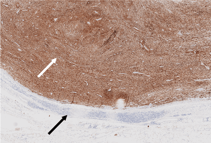
Tumor (white arrow) is diffusely positive for S100, supporting assumption of benign schwannoma; by contrast, malignant peripheral nerve sheath tumors only show focal S100 positivityadjacent capsule and lymphatic cuff (black arrow) show no immunoreactivity

Discussion

Adrenal incidentalomas are adrenal tumors discovered on an imaging study obtained for indications exclusive to adrenal conditions.1,2 A radiological pre-requisite for an adrenal tumor to be considered an incidentaloma is that the tumor must be greater than 1 cm in maximum diameter.3

The majority of resected incidentalomas are non-functioning cortical adenomas. Differential diagnoses can include benign diagnoses, such as nodular hyperplasia, myelolipoma, ganglioneuroma, hamartoma, hemangioma, leiomyoma, neurofibroma, and teratoma. However, potentially malignant neoplasms, such as pheochromocytoma and primary ACC, are high on differential diagnoses, especially in larger tumors with atypical imaging characteristics.1,4 In studies correlating radiological and pathological adrenal tumor characteristics, the prevalence of adrenal incidentalomas has been noted to be approximately 5 percent, with fewer than 10 percent demonstrating endocrine functionality and fewer than 5 percent malignant. Adrenocortical adenoma is the most common tumor type, with prevalence estimates ranging from 1.4 to 8.9 percent based on autopsy series.5,6 In another recent study, the prevalence of adrenal incidentalomas was noted to be four percent in middle-age and up to ten percent in the elderly. In 80 percent of the cases, the final diagnosis was a non-functional benign adenoma. Despite differences in the reported prevalence, the most common functional, although often asymptomatic, incidentaloma is pheochromocytoma, comprising 11–23 percent of incidental adrenal tumors.7

Schwannomas are benign nerve sheath tumors thought to arise from peripheral, motor, sympathetic, or cranial nerves of the head and neck region and upper and lower extremities. They are usually solitary tumors in the gastrointestinal tract derived from the Auerbach and the Meissner myenteric plexus. They are located primarily in the stomach as submucosal lesions, often challenging to differentiate from gastrointestinal stromal tumors.8,9 Schwannomas presenting in the adrenal gland, however, are quite uncommon. The innervation of the adrenal medulla is comprised of the phrenic nerve, the vagus nerve, and the sympathetic trunk, from which adrenal schwannomas are thought to arise.10 Malignant peripheral nerve sheath tumors (MPNSTs) are also very rare, comprising only 2 percent of all soft tissue sarcomas. MPNSTs are usually associated with neurofibromatosis type I syndrome and are almost always diagnosed on final microscopic histopathology and immunohistochemistry.11,12

To date, fewer than 50 cases of adrenal schwannomas have been reported in the literature. Almost all are non-functional tumors, except a single case report of an adrenal retroperitoneal schwannoma with catecholamine overproduction and oversecretion.13 Recognizing an adrenal schwannoma based on imaging features is very challenging, and the potential for malignancy is difficult to exclude. Thus, surgical resection is almost always the recommended approach, either open or laparoscopic, depending on size and on preoperative concerns for malignancy and surgeon preference.1,1416

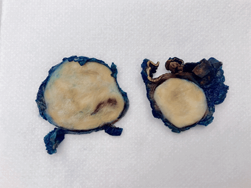
Gross and microscopic pathology of adrenal schwannoma typically reveal a firm, well-circumscribed rounded mass, tan-yellow to grayish-white in appearance, which can appear heterogeneous when larger in size (Figure 4). Adrenal schwannomas may or may not have a fibrous capsule but overall appear grossly similar to schwannomas originating from other sites. Immunohistochemistry stains are consistent with benign peripheral nerve sheath tumors, almost always with S-100 diffuse positivity, which differentiates these tumors from pheochromocytomas. Schwannomas are also characteristically negative for CD117, desmin, CD-34, HMB-45, synaptophysin, chromogranin, cytokeratin, and smooth muscle actin.10,14

Figure 4. Gross Photograph of Specimen, Showing Normal Grayish-White Appearance of Benign Schwannoma. Published with Permission

Current literature is limited regarding recommended follow-up of adrenal schwannomas. To our knowledge, there have been no reports of recurrence or metastatic disease reported in the literature.14,1719

The case presented here is unique. The patient presented with a non-functional left adrenal mass in the setting of radiologic mediastinal paraaortic lymphadenopathy, which on biopsy represented schwannoma and not lymphoid tissue. Additional pathologic analysis and multidisciplinary review concluded that both tumors represented benign schwannomas without evidence of invasive or malignant features. To our knowledge, only one other case of synchronous adrenal and mediastinal schwannoma has been reported in the literature. In this case, a patient presented with a paraspinal posterior mediastinal mass and left adrenal mass, both of which were managed with complete excision and were benign on final pathologic analysis.18 In our case, the decision was made to follow the mediastinal schwannomas with serial imaging based on its benign pathologic features and abutment of the descending thoracic aorta.

Conclusion

Adrenal schwannomas are a very rare entity, presenting almost always as an asymptomatic, non-functioning adrenal incidentaloma. Preoperative diagnosis cannot be clearly made by imaging characteristics and is almost always determined by final pathology. To date, there is no data to suggest a risk of recurrence or metastasis. The case presented here is unique in that this patient had a concurrent left adrenal incidentaloma and mediastinal para-aortic schwannoma, which raised concerns for possible metastatic disease. However, pathologic analysis demonstrated benign characteristics without evidence of an invasive component.

Lessons Learned

Benign adrenal schwannomas most commonly present as adrenal incidentaloma. Following biochemical evaluation, surgical resection remains the recommended treatment due to difficulty excluding adrenal malignancy based solely on preoperative radiological characteristics.

Authors

Mitsakos ATa, Stalls JSb; Parikh AAa; Snyder RAa,c

Author Affiliations

  1. Department of Surgery, Division of Surgical Oncology, Brody School of Medicine at East Carolina University, Greenville, NC 27834
  2. Department of Pathology and Laboratory Medicine, Brody School of Medicine at East Carolina University, Greenville, NC 27834
  3. Department of Public Health, East Carolina University, Greenville, NC 27834

Corresponding Author

Rebecca A. Snyder, MD, MPH, FACS
Brody School of Medicine at East Carolina University
600 Moye Boulevard, Ste. 4S-24
Greenville, NC 27834
Phone: (252) 744-4110
E-mail: snyderre19@ecu.edu

Disclosure Statement

The authors have no conflicts of interest to disclose.

Funding/Support

The authors have no financial relationships or in-kind support to disclose.

Received: January 6, 2020
Revision Revised: July 28, 2020
Accepted for Publication: October 29, 2020

References

  1. Linos DA. Adrenal incidentaloma (adrenaloma). Hormones (Athens). 2003;2(1):12-21. doi:10.14310/horm.2002.1178
  2. Farrugia FA, Martikos G, Surgeon C, et al. Radiology of the adrenal incidentalomas. Review of the literature. Endocr Regul. 2017;51(1):35-51. doi:10.1515/enr-2017-0005
  3. Papierska L, Cichocki A, Sankowski AJ, Cwikła JB. Adrenal incidentaloma imaging - the first steps in therapeutic management. Pol J Radiol. 2013;78(4):47-55. doi:10.12659/PJR.889541
  4. Mantero F, Terzolo M, Arnaldi G, et al. A survey on adrenal incidentaloma in Italy. Study Group on Adrenal Tumors of the Italian Society of Endocrinology. J Clin Endocrinol Metab. 2000;85(2):637-644. doi:10.1210/jcem.85.2.6372
  5. Blake MA, Cronin CG, Boland GW. Adrenal imaging [published correction appears in AJR Am J Roentgenol. 2012 May;198(5):1232]. AJR Am J Roentgenol. 2010;194(6):1450-1460. doi:10.2214/AJR.10.4547
  6. Lattin GE Jr, Sturgill ED, Tujo CA, et al. From the radiologic pathology archives: Adrenal tumors and tumor-like conditions in the adult: radiologic-pathologic correlation. Radiographics. 2014;34(3):805-829. doi:10.1148/rg.343130127
  7. Lee JM, Kim MK, Ko SH, et al. Clinical guidelines for the management of adrenal incidentaloma. Endocrinol Metab (Seoul). 2017;32(2):200-218. doi:10.3803/EnM.2017.32.2.200
  8. Melvin WS, Wilkinson MG. Gastric schwannoma. Clinical and pathologic considerations. Am Surg. 1993;59(5):293-296.
  9. Snyder RA, Harris E, Hansen EN, Merchant NB, Parikh AA. Gastric schwannoma. Am Surg. 2008;74(8):753-756.
  10. Mohiuddin Y, Gilliland MG. Adrenal schwannoma: a rare type of adrenal incidentaloma. Arch Pathol Lab Med. 2013;137(7):1009-1014. doi:10.5858/arpa.2012-0291-RS
  11. Farid M, Demicco EG, Garcia R, et al. Malignant peripheral nerve sheath tumors. Oncologist. 2014;19(2):193-201. doi:10.1634/theoncologist.2013-0328
  12. James AW, Shurell E, Singh A, Dry SM, Eilber FC. Malignant peripheral nerve sheath tumor. Surg Oncol Clin N Am. 2016;25(4):789-802. doi:10.1016/j.soc.2016.05.009
  13. Hou J, Zhang L, Guo Y, Chen H, Wang W. Primary adrenal schwannoma with catecholamine hypersecretion. Arch Med Sci. 2016;12(3):681-683. doi:10.5114/aoms.2016.59942
  14. Târcoveanu E, Dimofte G, Bradea C, et al. Adrenal schwannoma. JSLS. 2009;13(1):116-119.
  15. Abdessater M, El Mokdad M, Gas J, Sleiman W, Coloby P, Bart S. Juxta-adrenal schwannoma presenting as a giant adrenal tumor: A case report and a literature review. Int J Surg Case Rep. 2018;53:132-136. doi:10.1016/j.ijscr.2018.10.017
  16. Kumar S, Karthikeyan VS, Manohar CS, Sreelakshmi K, Shivalingaiah M. Adrenal schwannoma: a rare incidentaloma. J Clin Diagn Res. 2016;10(8):PD01-PD2. doi:10.7860/JCDR/2016/20405.8265
  17. Lau SK, Spagnolo DV, Weiss LM. Schwannoma of the adrenal gland: report of two cases. Am J Surg Pathol. 2006;30(5):630-634. doi:10.1097/01.pas.0000194739.80174.26
  18. Yang CY, Chou CW, Lin MB, Li CF. Schwannomas of the left adrenal gland and posterior mediastinum. J Chin Med Assoc. 2009;72(2):83-87. doi:10.1016/S1726-4901(09)70028-0
  19. Hsiao HL, Li CC, Lin HC, Yeh HC, Huang CH, Wu WJ. Adrenal schwannoma treated with laparoscopic adrenalectomy: a case report. Kaohsiung J Med Sci. 2008;24(10):553-557. doi:10.1016/S1607-551X(09)70016-6