Unsupported Browser
The American College of Surgeons website is not compatible with Internet Explorer 11, IE 11. For the best experience please update your browser.
Menu
Become a member and receive career-enhancing benefits

Our top priority is providing value to members. Your Member Services team is here to ensure you maximize your ACS member benefits, participate in College activities, and engage with your ACS colleagues. It's all here.

Become a Member
Become a member and receive career-enhancing benefits

Our top priority is providing value to members. Your Member Services team is here to ensure you maximize your ACS member benefits, participate in College activities, and engage with your ACS colleagues. It's all here.

Membership Benefits
ACS
Case Reviews in Surgery

Rare Care of Retrorectal Abscess and Meningocele

January 11, 2022

Abstract

Background

Retrorectal abscess associated with meningocele is very rare. Sacral pain and fever are common symptoms of retrorectal abscess. The significance of retrorectal abscess associated with pain, fever, and severe headache is presented in this unusual case presentation of retro rectal abscess and a pelvic meningocele.

Summary

A 41-year-old woman presented with fever, back pain in the sacral region, and headache resistant to painkillers of a two-week-long duration. The presence of a painful rectal mass on rectal examination prompted an abdominal MRI, which revealed a retrorectal inflamed mass compressing a meningocele with a hypoplastic dysplastic coccyx and last sacral elements. The patient was treated with IV antibiotics and surgical drainage of the retrorectal abscess through a sacral approach without compromising the meningocele. The meningocele compression was relieved and resulted in a decrease in intracranial pressure and headache. Three-year follow-up has revealed an asymptomatic meningocele with no recurrence of pelvic or cerebral problems.

Conclusion

Patient presenting with an inflamed rectal mass on rectal examination associated with fever and persistent headache should undergo pelvic MRI before surgery. This measure will assist the surgeon in planning a safe surgical approach to the pelvic abscess with a presence of a meningocele.

Key Words

retrorectal abscess; pelvic MRI; hypoplastic dysplastic coccyx; meningocele; retrorectal tumor; pelvic abscess; rectothecal fistula


Case Description

A 41-year-old woman presented with fever, back pain in the sacral region, and headache resistant to painkillers of a two-week-long duration. Past medical history was negative. On physical examination, there was an erythematous sacral perineal area painful to the touch (Figure 1).

Figure 1. Erythematous Sacral Area. Published with Permission

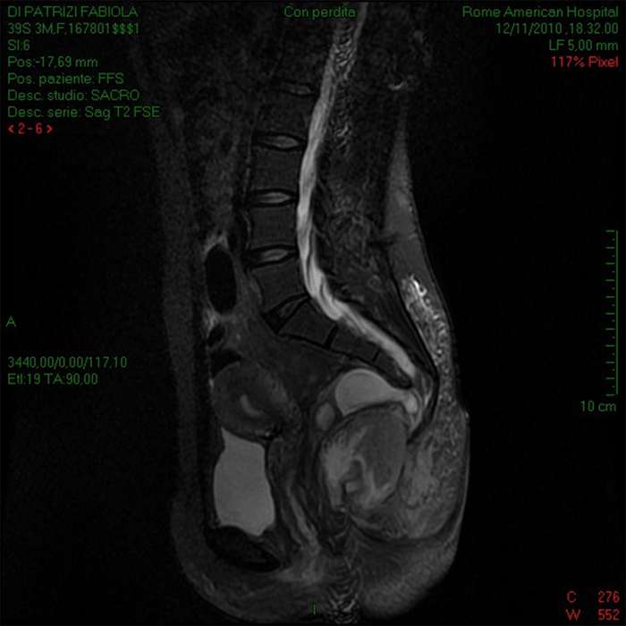
An anal exam revealed the presence of a painful posterior mass. Lab test: WBC 18.800, Hgb 10.1 g/dl ESR 100 mm/h. A pelvic sonogram indicated an 8.5 cm retrorectal mass without defined margins. An MRI of the abdomen was requested and showed the presence of presacral mass above the levator musculature, anterior to the retro sacral fascia, posterior to the rectum compressing a meningocele with a hypoplastic dysplastic coccyx and last sacral elements (Figure 2).

Figure 2. MRI Showing Presence of Retrorectal Presacral Mass Compressing Meningocele. Published with Permission

The patient was taken to the operating room, and a posterior sacral approach was chosen to drain the retrorectal abscess. Before skin incision, a left colonoscopy and reverse inspection of the anal canal was performed to exclude possible colonic or anal cryptoglandular disease as an origin of the pelvic abscess; methylene blue was injected in the colon and looked for when the abscess cavity was entered. Elliptical excision of skin in the area of the missing coccyx was made to guarantee large drainage (Figure 3).

Figure 3. Posterior Sacral Surgical Approach. Published with Permission

The retrorectal cavity was bluntly entered. There was no presence of methylene blue in the abscess cavity. The draining purulent material was sent for culture and cytology. The pus was totally evacuated, and the cavity was washed with saline solution—attention was paid to not touch the meningocele. Histology of the abscess wall revealed a diffuse inflamed necrotic wall. Antibiotic therapy was continued with ceftriaxone 2 g/day, metronidazole 1.5 g/day, and amikacin 1 g/day. Escherichia coli was cultured from the abscess cavity. The patient was discharged after ten days without pain, fever, or headache. Follow up at 3, 6, 12, 24, and 36 months revealed an asymptomatic patient (Figure 4).

Figure 4. Follow-up MRI Showing Resolution of Retrorectal Abscess and Expansion of Meningocele. Published with Permission

Discussion

Retrorectal abscess associated with meningocele is very rare.13 Meningocele is a cystic meningeal protrusion through the presacral space. Anterior meningocele can be related to some agenesis of the anterior sacrum. When associated with congenital caudal anomalies, anal stenosis and sacral defect can be part of the Currarino syndrome.46 Epidermoid and dermoid cysts represent 50 percent of retrorectal mass and result from defective closure of ectodermal tube; 30 percent of these cysts can become infected form an abscess with or without fistula, and are benign.

Complications of a meningocele are headache, infection with or without fistulization, rectothecal fistula, and meningitis. The symptoms of retrorectal tumors depend on the size of the mass and location. Sacral pain is a common symptom associated with fever in case of an abscess or urinary infection.7 The significance of retrorectal abscess associated with pain, fever, and severe persistent headache is presented in this unusual case presentation of retrorectal abscess and a pelvic meningocele. Pappalardo and coauthors propose a preoperative CT and an MRI anatomical and topographical classification to allow an adequate surgical approach.8

Our surgical strategy included preoperative colonoscopy to exclude possible colonic or anal origin for the abscess. This measure allowed us to approach the retrorectal abscess through a posterior incision in the coccygeal area, avoiding meningocele injury. The meningocele was asymptomatic for three years; therefore, no further surgical treatment was deemed necessary by our neurosurgeons. The histology of the abscess wall revealed an inflamed necrotic tissue; there was no confirmation of a suspected cystic origin infection. For this reason, the patient was followed up with an MRI for three years with negative radiological findings.

Conclusion

A patient with a rectal abscess mass on rectal examination associated with fever and persistent headache should undergo an MRI before surgery. This approach will help the surgeon evaluate the extent and source of the abscess and exclude the presence of a meningocele to plan a proper surgical approach and avoid possible meningitis and meningo-perineal fistula from the surgery.

Lessons Learned

When a rectal abscess mass is found on rectal examination associated with fever and persistent headache, sacral meningocele is a possibility. MRI will confirm the presence of a meningocele, assisting the surgeon in planning a procedure to avoid possible meningitis and meningo-perineal fistula.

References

  1. Jao SW, Beart RW Jr, Spencer RJ, Reiman HM, Ilstrup DM. Retrorectal tumors. Mayo Clinic experience, 1960-1979. Dis Colon Rectum. 1985;28(9):644-652. doi:10.1007/BF02553440
  2. Jackman RJ, Clark PL, Smith ND. Retrorectal tumors. JAMA. 1951;145(13):956–962. doi:10.1001/jama.1951.02920310012003
  3. Buyukbese Sarsu S, Parmaksiz ME, Cabalar E, Karapur A, Kaya C. A very rare cause of anal atresia: Currarino syndrome. J Clin Med Res. 2016;8(5):420-423. doi:10.14740/jocmr2505w
  4. Lynch SA, Wang Y, Strachan T, Burn J, Lindsay S. Autosomal dominant sacral agenesis: Currarino syndrome. J Med Genet. 2000;37(8):561-566. doi:10.1136/jmg.37.8.561
  5. Dahan H, Arrivé L, Wendum D, Docou le Pointe H, Djouhri H, Tubiana JM. Retrorectal developmental cysts in adults: clinical and radiologic-histopathologic review, differential diagnosis, and treatment. Radiographics. 2001;21(3):575-584. doi:10.1148/radiographics.21.3.g01ma13575
  6. Uhlig BE, Johnson RL. Presacral tumors and cysts in adults. Dis Colon Rectum. 1975;18(7):581-589. doi:10.1007/BF02587141
  7. Pappalardo G, Frattaroli FM, Casciani E, et al. Retrorectal tumors: the choice of surgical approach based on a new classification. Am Surg. 2009;75(3):240-248.

Authors

Antonellis Fa; Falasco Ga; Trentino Pb; Ravo Bc

Author Affiliations

  1. Department of Surgery, Rome American Hospital, Rome, Italy
  2. San Camillo Forlanini Hospital, Università La Sapienza, Rome, Italy
  3. San Camillo Forlanini Hospital, Università Campus Biomedico, Rome, Italy

Corresponding Author

Biagio Ravo, MD, FACS
San Camillo Forlanini Hospital, Università Campus Biomedico
Via Emilio Longoni, 81, 00155
Rome, Italy

Disclosure Statement

The authors have no conflicts of interest to disclose.

Funding/Support

The authors have no relevant financial relationships or in-kind support to disclose.

Meeting Presentation

ACS Italian Chapter Congress, Convention Center La Nuvola 14‒18, Rome, Italy, October 2018

Received: November 9, 2020
Revision received: May 17, 2021
Accepted: October 21, 2021