Unsupported Browser
The American College of Surgeons website is not compatible with Internet Explorer 11, IE 11. For the best experience please update your browser.
Menu
Become a member and receive career-enhancing benefits

Our top priority is providing value to members. Your Member Services team is here to ensure you maximize your ACS member benefits, participate in College activities, and engage with your ACS colleagues. It's all here.

Become a Member
Become a member and receive career-enhancing benefits

Our top priority is providing value to members. Your Member Services team is here to ensure you maximize your ACS member benefits, participate in College activities, and engage with your ACS colleagues. It's all here.

Become a Member
ACS
ACS Case Reviews

Duodenal Atresia, Intestinal Malrotation, and Gastrojejunal Intussusception: Trisomy 21 with Complications

December 16, 2022

Abstract

Background

Gastrojejunal intussusception occurs as a rare complication following gastrojejunostomy.

Summary

A 50-year-old female with Down syndrome presented to the hospital with abdominal pain and multiple episodes of nonbilious emesis over the past 24 hours. She had a midline abdominal incision from prior surgery as an infant without any known historic details available. She was clinically stable, and the initial radiographic workup demonstrated an intussusception of jejunum into the stomach and signs of intestinal malrotation. Further investigation with upper endoscopy revealed the presence of complete duodenal atresia and viable jejunum within the gastric lumen. Preoperative radiologic evaluation and endoscopy suggested the intraoperative findings noted. We further describe this entity’s unique surgical management and briefly discuss the pertinent literature.

Conclusion

Optimal surgical management of gastrojejunal intussusception is dependent upon operative findings and bowel viability.

Key Words

gastrojejunal intussusception; gastrojejunostomy; trisomy 21, intestinal malrotation; duodenal atresia


Case Description

A 50-year-old noncommunicative female with Down syndrome, hypothyroidism, and severe developmental delay presented to the emergency room with abdominal pain and multiple episodes of nonbilious emesis over the past 24 hours. There was a history of a congenital cardiac defect as well. She was mildly distended and tender on physical examination without signs of peritonitis. There was a midline abdominal scar and no external hernias. The rectal exam was Hemoccult negative. Laboratory analysis revealed a white blood cell count of 13,000 and normal serum chemistries.

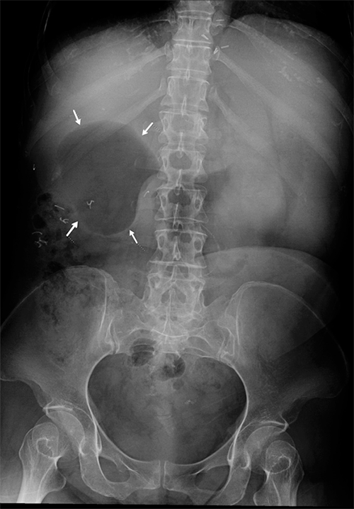
The patient’s initial radiographic workup consisted of a plain abdominal film demonstrating a large, gas-filled structure in the right upper quadrant (Figure 1). Careful analysis of a subsequent CT scan of the abdomen and pelvis with oral but no IV contrast demonstrated several findings, the first and foremost of which was what appeared to be an intussusception of the small bowel into the stomach. Upon further analysis of the CT scan, it appeared the small bowel was located predominantly on the right side of the abdomen while the colon occupied the left. While challenging to discern adequately without IV contrast, the SMV was in front of the SMA, but the two vessels were not fully transposed. An additional finding was that of contrast opacification in the right upper quadrant in the same area where a gas-filled loop of bowel had been noted on the initial plain film. Finally, the duodenum did not appear to cross the midline abdomen (Figure 2).

Since the patient was stable, consent was obtained for an initial upper endoscopy to further define and clarify anatomy, followed by laparotomy immediately thereafter. The operating surgeon had full anesthesia capabilities and performed the endoscopy in our surgical endoscopy unit. Upon passage of the scope into the distal gastric body, an intussuscepting limb of the small bowel was encountered, which was fully viable. Its origin was not initially apparent, and the scope could be maneuvered through the patient’s native pylorus into a large blind pouch of proximal duodenum with no outlet. There was no bile in the stomach or this blind ending pouch (Figure 3). A laparotomy followed, and examination of the abdominal content revealed the colon to indeed be on the left side of the abdomen. The appendix was surgically absent, consistent with prior Ladd’s procedure. There was complete atresia of the second portion of the duodenum. A surgical gastrojejunostomy was noted, the efferent limb of which had intussuscepted retrograde through the gastrojejunal anastomosis into the gastric lumen. The intussusception was manually reduced by grasping the stomach at the cephalad aspect of the intussusception and pushing the jejunum within its lumen distally back toward the anastomosis while placing only minimal traction on the intussuscepted jejunal limb from below. Once fully reduced, there was no evidence of a lead point, and jejunal viability was confirmed (Figure 4).

A decision was made to pexy the efferent anastomotic limb further along the greater curve of the stomach and to an adjacent portion of jejunum immediately beyond to prevent future intussusception episodes (Figure 5). The efferent limb pexy was created with a gentle curve in mind to avoid possible kinking or obstruction at its apex. Postoperative GI contrast studies and a CT scan confirmed normal transit through this area (Figure 6). Postoperatively, the patient did well and recovered to discharge several days later.

Figure 1. Initial Abdominal X ray Demonstrating Large Proximal Duodenal Gas Bubble. Published with Permission

Figure 2. CT Imaging. Published with Permission

Oral contrast demonstrates findings of intussusception (right); intussusceptum (jejunum) is seen within intussuscipiens (body of stomach). Oral contrast is identified within stomach and blind-ending proximal duodenum.

Figure 3. Endoscopic Images. Published with Permission

Noted viable intussucepted jejunum in gastric antrum (left) and blind-ending proximal duodenal pouch (right).

Figure 4. Intraoperative Images. Published with Permission

Jejunogastric intussusception (left) and post-reduction viable small bowel (right) are shown.

Figure 5. Diagram of Post-Pexy Surgical Anatomy

Figure 6. Postoperative Upper Gastrointestinal Series and CT. Published with Permission

Image demonstrates persistently dilated and air-filled duodenal bulb in right upper quadrant and right-sided small bowel opacification with contrast.

Discussion

Down syndrome is associated with various gastrointestinal anomalies, including duodenal atresia (6%),1 annular pancreas, gastrointestinal reflux, imperforate anus, and Hirschsprung disease. Additionally, malrotation, Meckel’s diverticulum, and duodenal web appear more common in this population.2‒5 Congenital cardiac defects are also well described in these patients. They may be a potential source of surgical morbidity.6 Current operative management of duodenal atresia consists of duodenoduodenostomy or duodenojejunostomy.7 Perhaps at the time of initial presentation and exploration as an infant in our patient, it may not have been precisely clear where the bile and pancreatic ducts entered the atretic duodenum and so gastrojejunostomy may have been chosen instead.

Jejunogastric intussusception following gastrojejunostomy is exceedingly rare, with approximately 300 cases reported in the literature. This finding arises as a complication of prior surgical anastomosis. Most reported cases (74%) involve the efferent limb of the gastrojejunostomy, but afferent and combined limb intussusception have been described.8‒10 The mechanism is likely related to retrograde peristalsis. Still, long afferent limb, jejunal spasm, abnormal motility, hyperacidity, and increased intraabdominal pressure have also been implicated.11,12 There were no lead points for this condition described in the literature, so resection based solely on concern for anatomic causes appears unnecessary.

Endoscopic reduction has been described, but the risk of perforation or recurrence is high.13,14 Potential surgical options for intussusception depend largely upon intraoperative findings and intestinal viability. They include simple reduction with or without fixation and revision or resection with re anastomosis.8 Another potential option is to convert the gastrojejunostomy to Roux-en-Y configuration, mainly if nonviable intestine is encountered at exploration. In our case, the afferent limb was short in length because the index gastrojejunostomy had been constructed close to the duodenum, completely to the right of the midline. The limb was also partially tethered in the retroperitoneum from prior surgery. If a long afferent limb had been encountered, additional pexy of the afferent to the efferent limb, similar to a Braun jejujo-jejunostomy but without anastomosis, could have also been considered.

Conclusion

We report a case of gastrojejunal intussusception, an exceedingly rare complication of gastrojejunostomy with a good outcome. Optimal surgical management is dependent upon operative findings and bowel viability.

Lessons Learned

Careful analysis of preoperative imaging is especially useful when no prior surgical historical details are available. Endoscopy can be useful, especially when gastrojejunal intussusception is suggested by radiologic workup in a stable patient. The lack of a lead point in all reported cases makes resection unnecessary unless bowel viability is in question.

Acknowledgments

Dr. Martínez-Zavala and Mariana Tapia Vazquez contributed to the illustration.

References

  1. Stoll C, Alembik Y, Dott B, Roth MP. Study of Down syndrome in 238,942 consecutive births. Ann Genet. 1998;41(1):44-51.
  2. Eksarko P, Nazir S, Kessler E, et al. Duodenal web associated with malrotation and review of literature. J Surg Case Rep. 2013;2013(12):rjt110. Published 2013 Dec 18. doi:10.1093/jscr/rjt110
  3. Mirza B, Sheikh A. Multiple associated anomalies in patients of duodenal atresia: a case series. J Neonatal Surg. 2012;1(2):23. Published 2012 Apr 1.
  4. Frid C, Drott P, Lundell B, Rasmussen F, Annerén G. Mortality in Down's syndrome in relation to congenital malformations. J Intellect Disabil Res. 1999;43 ( Pt 3):234-241. doi:10.1046/j.1365-2788.1999.00198.x
  5. Buchin PJ, Levy JS, Schullinger JN. Down's syndrome and the gastrointestinal tract. J Clin Gastroenterol. 1986;8(2):111-114. doi:10.1097/00004836-198604000-00002
  6. Angotti R, Molinaro F, Sica M, et al. Association of Duodenal Atresia, Malrotation, and Atrial Septal Defect in a Down-Syndrome Patient. APSP J Case Rep. 2016;7(2):16. Published 2016 Apr 24.
  7. Fonkalsrud EW, DeLorimier AA, Hays DM. Congenital atresia and stenosis of the duodenum. A review compiled from the members of the Surgical Section of the American Academy of Pediatrics. Pediatrics. 1969;43(1):79-83.
  8. Irons HS Jr, Lipin RJ. Jejuno-gastric intussusception following gastro-enterostomy and vagotomy. Ann Surg. 1955;141(4):541-546. doi:10.1097/00000658-195504000-00017
  9. Rather SA, Dar TI, Wani RA, Khan A. Jejunogastric intussusception presenting as tumor bleed. J Emerg Trauma Shock. 2010;3(4):406-408. doi:10.4103/0974-2700.70775
  10. Czerniak A, Bass A, Bat L, Shemesh E, Avigad I, Wolfstein I. Jejunogastric intussusception. A new diagnostic test. Arch Surg. 1987;122(10):1190-1192. doi:10.1001/archsurg.1987.01400220100019
  11. Waits JO, Beart RW Jr, Charboneau JW. Jejunogastric intussusception. Arch Surg. 1980;115(12):1449-1452. doi:10.1001/archsurg.1980.01380120023006
  12. Archimandritis AJ, Hatzopoulos N, Hatzinikolaou P, et al. Jejunogastric intussusception presented with hematemesis: a case presentation and review of the literature. BMC Gastroenterol. 2001;1:1. doi:10.1186/1471-230x-1-1
  13. Kochhar R, Saxena R, Nagi B, Gupta NM, Mehta SK. Endoscopic management of retrograde jejunogastric intussusception. Gastrointest Endosc. 1988;34(1):56-57. doi:10.1016/s0016-5107(88)71233-x
  14. Toth E, Arvidsson S, Thorlacius H. Endoscopic reduction of a jejunogastric intussusception. Endoscopy. 2011;43 Suppl 2 UCTN:E63. doi:10.1055/s-0030-1256103

Authors

Martínez-Zavala VEa; Cataneo-Serrato Ja; Dwijendra STb, Major SMb; Hyser MJc

Author Affiliations

  1. University of Illinois College of Medicine at Chicago (Metropolitan Group), Chicago, IL 60657
  2. Department of Diagnostic Radiology, AMITA Health Saint Francis Hospital, Evanston, IL 60202
  3. Department of Surgery, AMITA Health Saint Francis Hospital, Evanston, IL 60202

Corresponding Author

Victor E. Martínez-Zavala, MD
Advocate Illinois Masonic Medical Center
836 W. Wellington Avenue, Ste. 4800
Chicago, IL 60657
Phone: 913-601-1972
Email: vemz.1989@gmail.com

Disclosure Statement

The authors have no conflicts of interest to disclose.

Funding/Support

The authors have no relevant financial relationships or in-kind support to disclose.

Received: September 8, 2020
Revision received: November 8, 2020
Accepted: November 24, 2020