Abstract
Background
We present a 78-year-old female with small bowel obstruction due to a jejunal mass. The patient underwent an exploratory laparotomy finding a small bowel obstruction due to a tumor in the mid-jejunum. We performed a segmental bowel resection, including the mass. Final pathology of the mass was consistent with gastrointestinal schwannoma.
Summary
This patient presented with acute onset of diffuse abdominal pain, nausea, and emesis. A CT scan of the abdomen and pelvis and CT Enterography revealed a jejunal mass causing a partial blockage. The patient underwent an exploratory laparotomy with segmental jejunal resection. Final pathology was consistent with a gastrointestinal schwannoma.
Schwannomas are slow-growing mesenchymal tumors that rarely occur in the gastrointestinal tract. They represent a rare cause of submucosal masses found in the GI tract and are commonly diagnosed postoperatively given their indolent clinical course and often nonspecific imaging findings. The most common site of gastrointestinal schwannomas is the stomach. Macroscopically they are indistinguishable from the much more common GIST tumor. Moreover, neither imaging nor endoscopic appearance is pathognomonic. The diagnosis is usually secured after surgical resection and immunohistochemical staining.
Conclusion
Small gastrointestinal schwannomas are difficult to diagnose preoperatively. We present a case with a rare location of a gastrointestinal schwannoma leading to the uncommon presentation with a small bowel obstruction. Due to their excellent prognosis following resection, schwannomas are important to differentiate from other malignancies with similar presentation.
Key Words
jejunal schwannoma; mesenchymal small bowel tumors; small bowel obstruction
Case Description
This case review discusses the disease presentation and further course of a 78-year-old female. The patient initially presented to the emergency department in November 2020. At that time, she complained of abdominal discomfort with rapidly progressive nausea and emesis over the course of the day. She reported numerous episodes of bilious emesis and epigastric and upper abdominal discomfort, similar to her prior experienced episode of pancreatitis.
The patient's past medical history was fairly unremarkable, with controlled hypertension and hypothyroidism. The patient experienced one episode of pancreatitis over 20 years ago following a cholecystectomy. Her remaining surgical history includes a total abdominal hysterectomy in the 1990s and an orthopedic surgery for a right humerus fracture. The patient's family history was relevant for pancreatic adenocarcinoma in her father.
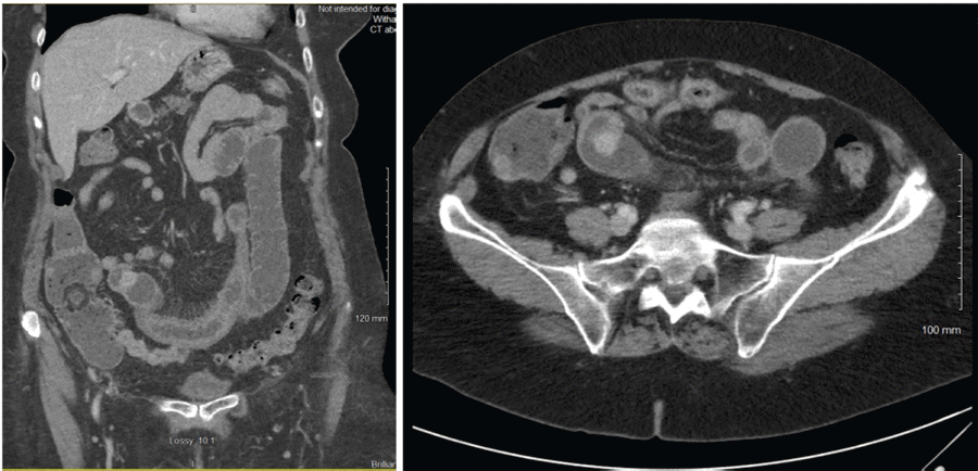
The patient's workup in the emergency department included a CBC, CMP, and lipase with no relevant findings. A CT scan of her abdomen and pelvis with intravenous contrast with 5 mm slides a small bowel mass in the right lower quadrant causing a small bowel obstruction (Figure 1). The patient improved after symptomatic treatment in the emergency department and was released with follow-up with a gastroenterologist. In the following weeks, she underwent a CT enterography that showed extensive diverticula and a 1.3 cm enhancing mass versus focal peristalsis in the jejunum. A subsequent colonoscopy in late January showed pancolonic diverticulosis and two small tubular adenomas but was otherwise unremarkable. She was recommended to follow up with a surgeon, given the location of the mass, presumably in the mid-small bowel and therefore out of reach for most endoscopic practices.
The patient was taken to the operating room in late February, and a lower midline laparotomy was performed with segmental bowel resection. The patient was noted to have sparse adhesions, likely related to her prior abdominal surgeries. Her small bowel was eviscerated and inspected from the ligament of Treitz to the ileocecal valve. A hard, intramural mass was discovered at approximately the mid-jejunal location. This was believed to be the most likely cause of her prior obstructive episode. A segmental resection with stapled anastomosis was performed. The specimen was opened on a back table, and a nodular submucosal mass was noted. As mentioned above, the macroscopic morphology was not distinct for a schwannoma. The patient tolerated the procedure well and was discharged on postoperative day 4. The patient reported resolution of her abdominal symptoms at follow-up in clinic two weeks postoperatively and via phone at four weeks. She experienced no further obstructive episodes, and her abdominal discomfort had resolved.
Figure 1. CT of Abdomen and Pelvis with IV Contrast Showing 1.2 cm Mass in Mid-Jejunum. Published with Permission
The resection specimen was examined by pathology. It showed a 1.5 cm tumor with involvement of the serosal surface in a 5.2 cm long jejunal segment. The tumor was macroscopically gray-white and indurated. Histological examination was consistent with a benign nerve sheath tumor with negative margins negative and six benign lymph nodes in the specimen. Closer examination showed a submucosal spindle cell neoplasm surrounded by bands of lymphoid tissue. Mitotic figures were rare to absent.
Immunohistochemical workup showed focal AE1-3 and strongly positive S100 staining. Further stains for DOG1, CD117, SMA, Melan-A, and CAM5.2 were negative. Ki-67 showed a low proliferation rate. The tumor was diagnosed as schwannoma based on gross microscopic findings and immunohistochemical workup.
Discussion
Schwannomas are neoplasms arising from myelin sheath-producing cells of the peripheral nerves. A gastrointestinal location of schwannomas is rare and reported for approximately 2-6% of all schwannomas.1 The most common gastrointestinal location is the stomach; here, schwannomas represent about 4% of all benign gastric tumors.2,3 The second most common GI location of schwannomas is the colon and rectum. Schwannomas of the small intestine are exceedingly rare.4
Schwannomas tend to be benign but can cause neural dysfunction. This is well demonstrated by their common presenting symptom of hearing loss and vertigo in cases of cochleovestibular tumors.5,6 Most schwannomas are considered sporadic, even though a genetic disposition in patients with type II neurofibromatosis has been described.7,8 Clinical presentation of gastrointestinal schwannomas may be, as in this case, with obstructive symptoms; more commonly, they are asymptomatic and incidentally found on imaging or during surgery.9
They tend to show a homogenous pattern of tumor attenuation if they can be radiographically visualized. They may appear primarily intraluminal, extraluminal, or both without clearly defined enhancement.10 Smaller-sized gastrointestinal stromal tumors that lack hemorrhagic or cystic components may show a similar CT appearance.11,12
The difficulty of preoperative diagnosis based on imaging alone becomes especially apparent with esophageal tumor locations. A much more common intramural leiomyoma is indistinguishable from an esophageal schwannoma on CT scan.13,14
Given the difficulty of diagnosis based on imaging findings alone, the pathology is crucial in diagnosing intestinal schwannomas. As seen in this case, gastrointestinal schwannomas commonly have a low mitotic count with characteristic peripheral lymphoid cuff.15,16 Immunohistochemical staining typically shows a strongly positive reaction to S-100 protein, while CD117, smooth muscle actin, and desmin are negative, which differentiates these tumors from the gastrointestinal stromal tumors and leiomyomas, respectively.4,16
Further preoperative workup of suspected small bowel schwannomas with endoscopy or CT enterography is unlikely to yield additional information. Tumors of the small intestine should undergo surgical exploration and excision if feasible.17,18 The treatment of choice for gastrointestinal schwannomas is resection.4,19 This secures the diagnosis and provides an excellent prognosis.15
Conclusion
This case demonstrates the presence of a gastrointestinal schwannoma and the difficulty with diagnosis, especially when located in the small bowel. This patient underwent multiple noninvasive and invasive tests, which were able to demonstrate the jejunal mass but were insufficient to establish a diagnosis. Further workup could have been obtained with capsule endoscopy and possibly even push-pull enteroscopy. Still, given the submucosal location of these tumors, it is possible no additional information would have been provided. This leads one to the conclusion that surgical resection and immunohistochemical workup is the most promising route to securing a diagnosis and providing adequate treatment simultaneously in these rare cases.
Lessons Learned
Schwannomas are a rare entity in the gastrointestinal tract but are an important differential diagnosis given their benign clinical course following resection. The diagnosis remains difficult to secure prior to resection.
Authors
Lueders A; Saxe J; Kaderabek D
Author Affiliation
Ascension St. Vincent Hospital Indianapolis, Indianapolis, IN 46260
Corresponding Author
Amelie Lueders, MD
2001 W. 86th Street
Indianapolis, IN 46260
Email: ami.lueders@outlook.com
Disclosure Statement
The authors have no conflicts of interest to disclose.
Funding/Support
The authors have no relevant financial relationships or in-kind support to disclose.
Received: April 2, 2021
Revision received: July 6, 2021
Accepted: July 27, 2021
References
- Wilde BK, Senger JL, Kanthan R. Gastrointestinal schwannoma: an unusual colonic lesion mimicking adenocarcinoma. Can J Gastroenterol. 2010;24(4):233-236. doi:10.1155/2010/943270
- Lin CS, Hsu HS, Tsai CH, Li WY, Huang MH. Gastric schwannoma. J Chin Med Assoc. 2004;67(11):583-586.
- Zheng L, Wu X, Kreis ME, et al. Clinicopathological and immunohistochemical characterisation of gastric schwannomas in 29 cases. Gastroenterol Res Pract. 2014;2014:202960. doi:10.1155/2014/202960
- Miettinen M, Shekitka KM, Sobin LH. Schwannomas in the colon and rectum: a clinicopathologic and immunohistochemical study of 20 cases. Am J Surg Pathol. 2001;25(7):846-855. doi:10.1097/00000478-200107000-00002
- Roosli C, Linthicum FH Jr, Cureoglu S, Merchant SN. Dysfunction of the cochlea contributing to hearing loss in acoustic neuromas: an underappreciated entity. Otol Neurotol. 2012;33(3):473-480. doi:10.1097/MAO.0b013e318248ee02
- von Kirschbaum C, Gürkov R. Audiovestibular Function Deficits in Vestibular Schwannoma. Biomed Res Int. 2016;2016:4980562. doi:10.1155/2016/4980562
- Evans DG, Birch JM, Ramsden RT. Paediatric presentation of type 2 neurofibromatosis. Arch Dis Child. 1999;81(6):496-499. doi:10.1136/adc.81.6.496
- Kawsar KA, Haque MR, Chowdhury FH. Abdominal schwannoma in a case of neurofibromatosis type 2: A report of a rare combination. Asian J Neurosurg. 2017;12(1):89-91. doi:10.4103/1793-5482.145347
- Mekras A, Krenn V, Perrakis A, et al. Gastrointestinal schwannomas: a rare but important differential diagnosis of mesenchymal tumors of gastrointestinal tract. BMC Surg. 2018;18(1):47. Published 2018 Jul 25. doi:10.1186/s12893-018-0379-2
- Levy AD, Quiles AM, Miettinen M, Sobin LH. Gastrointestinal schwannomas: CT features with clinicopathologic correlation. AJR Am J Roentgenol. 2005;184(3):797-802. doi:10.2214/ajr.184.3.01840797
- Burkill GJ, Badran M, Al-Muderis O, et al. Malignant gastrointestinal stromal tumor: distribution, imaging features, and pattern of metastatic spread. Radiology. 2003;226(2):527-532. doi:10.1148/radiol.2262011880
- Levy AD, Remotti HE, Thompson WM, Sobin LH, Miettinen M. Gastrointestinal stromal tumors: radiologic features with pathologic correlation. Radiographics. 2003;23(2):283-532. doi:10.1148/rg.232025146
- Matsuki A, Kosugi S, Kanda T, et al. Schwannoma of the esophagus: a case exhibiting high 18F-fluorodeoxyglucose uptake in positron emission tomography imaging. Dis Esophagus. 2009;22(4):E6-E10. doi:10.1111/j.1442-2050.2007.00712.x
- Yang PS, Lee KS, Lee SJ, et al. Esophageal leiomyoma: radiologic findings in 12 patients. Korean J Radiol. 2001;2(3):132-137. doi:10.3348/kjr.2001.2.3.132
- Kwon MS, Lee SS, Ahn GH. Schwannomas of the gastrointestinal tract: clinicopathological features of 12 cases including a case of esophageal tumor compared with those of gastrointestinal stromal tumors and leiomyomas of the gastrointestinal tract. Pathol Res Pract. 2002;198(9):605-613. doi:10.1078/0344-0338-00309
- Daimaru Y, Kido H, Hashimoto H, Enjoji M. Benign schwannoma of the gastrointestinal tract: a clinicopathologic and immunohistochemical study. Hum Pathol. 1988;19(3):257-264. doi:10.1016/s0046-8177(88)80518-5
- Lomdo M, Setti K, Oukabli M, Moujahid M, Bounaim A. Gastric schwannoma: a diagnosis that should be known in 2019. J Surg Case Rep. 2020;2020(1):rjz382. Published 2020 Jan 13. doi:10.1093/jscr/rjz382
- Ashley SW, Wells SA Jr.v Tumors of the small intestine. Semin Oncol. 1988;15(2):116-128.
- Fukushima N, Aoki H, Fukazawa N, Ogawa M, Yoshida K, Yanaga K. Schwannoma of the Small Intestine. Case Rep Gastroenterol. 2019;13(2):294-298. Published 2019 Jun 28. doi:10.1159/000501065

