Abstract
Background
A 59-year-old male was incidentally found to have a granular cell tumor of the cecum.
Summary
Granular cell tumors (GCTs) are uncommon soft tissue neoplasms that may present in various organs throughout the body. The most common locations are the subcutaneous tissue and the oral cavity; however, they have been reported in the submucosa of the gastrointestinal tract (GIT) from the esophagus to the rectum. GCTs are often found incidentally on routine endoscopy, and unlike most submucosal lesions, they possess malignant potential. We report a case of a GCT found in the cecum on a routine endoscopy of a 59-year-old asymptomatic male.
Conclusion
GCTs of the GIT are rare but require attention due to their malignant potential. We present a case of a GCT in the cecum that required surgical resection. This case report highlights the incidence of GCTs involving the GIT, the necessity of resection, and the different options for treatment.
Key Words
granular cell tumor; cecal tumors; cecal granular cell tumor
Case Description
The authors present a case of a rare colonic lesion requiring surgical resection for a granular cell tumor. The patient is a 59-year-old, asymptomatic male who was found to have a submucosal cecal mass on routine colonoscopy, which the endoscopist felt uncomfortable removing. There was no remarkable past medical or surgical history, but he was a 30 pack-year smoker. He had a significant family history of breast cancer in his mother and sister.
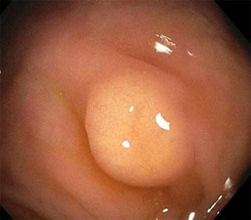
On colonoscopy, a solitary nodular lesion was found in the cecum with yellow discoloration, suggestive of a lipoma on gross examination (Figure 1). Gross appearance was lipomatous with a positive "pillow" sign. Biopsies were performed, and histology of the lesion revealed a granular cell tumor with IHC stains positive for S100, Calretinin, PAS, and CD68 and negative for AE1/3, CK7, CK20, Vimentin, and Desmin.
Figure 1. Solitary Submucosal Lesion in Cecum Seen on Colonoscopy. Published with Permission
The endoscopist felt the lesion was not amenable to endoscopic resection given the location and multiple failed attempts and referred the patient for surgical resection. The patient underwent a laparoscopic right hemicolectomy with an uneventful postoperative course.
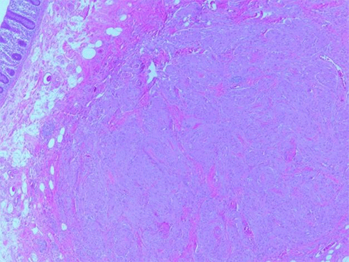
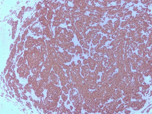
Final anatomic pathology revealed the following: 5 × 3 mm granular cell tumor of the submucosa at the cecum without invasion of the muscularis propria (Figure 2), IHC staining positive for S100 (Figure 3A) and CD68 (Figure 3B), 28 reactive lymph nodes without evidence of metastasis. Small bowel, colonic, and soft tissue margins were tumor-free.
Figure 2. Two H&E Stains. Published with Permission
Figure 3. Immunohistochemical Staining of Tumor Showing Positive Immunoreaction for S100 Protein and CD68
Discussion
Abrikossoff first described GCTs in 1926 as a form of muscle tumor.2,4 GCTs likely result from the proliferation of Schwann-like cells and histologically have a coarse, eosinophilic appearance with positive periodic acid–Schiff (PAS) staining.3,4 The granular nature of the tumor cells is due to the accumulation of secondary lysosomes in the cytoplasm.4 Typically, the overwhelming majority are solitary nodules.1,2,4 GCTs most commonly (45% to 65%) manifest in the head and neck region, specifically in the oral cavity, skin, and subcutaneous tissue.2,3,9 Involvement of the GIT is rare and has only been seen in about 1% to 11% of reported cases, with one-third of these occurring in the esophagus.4,5,8,9
GCTs are generally benign; however, 1% to 3% of identified lesions have been reported as malignant.5,9 When malignant, they are often larger than 5 cm, margins are unclear with surrounding tissue, invade the fat and/or muscle tissue, and are strongly positive for S100 and neuron-specific enolase on immunohistochemistry (IHC).5 Neither biopsies nor EUS can reliably distinguish benign GCT from malignant.5 Taking the malignant potential into consideration, the management of GCTs requires prompt excision via endoscopy or surgical resection if endoscopic resection is not feasible. Limited data is available regarding the rate of lymph node metastasis of GCTs of the colon owed to its rarity. A case series of 98 cases revealed a 1% chance of lymph node metastasis.10 Laparoscopic cecectomy was considered; however, the surgeon felt a formal oncological laparoscopic right hemicolectomy was the preferred operation due to the lack of data available regarding these rare tumors in the colon.
This patient's GCT is unusual, given its location. As stated, GCTs usually occur in the tongue and the skin, while 1% to 11% of GCTs occur in the GI tract.4,5,8,9 Of those occurring in the GIT, the majority are found in the esophagus, duodenum, anus, or stomach.4,5,8,9 Rarely are GCTs found in the colon or rectum.4,5,8,9 Since the discovery of GCTs, 130 cases of colonic GCTs have been identified in English-language journals.9 Colonic GCTs are most commonly found incidentally on screening colonoscopy, such as in this case, or when investigating other suspected lesions.5,7-9
The diagnosis of GCT depends on histopathology with a variable architecture ranging from small and well-circumcised nodules to larger and poorly circumscribed lesions.2,3 They may sometimes display an infiltrative property with satellite nodules as well.2,3 GCT cells are typically rounded epithelioid with a diffusely granular eosinophilic cytoplasm (Figure 3). The cytoplasm of the tumor cells accumulates secondary lysosomes responsible for the granular appearance.4 The lysosomes stain with PAS and are diastase resistant.4 IHC positivity for CD68 and neural marker S100 supports the diagnosis of GCT.2,4,9 Despite a large amount of IHC studies, the histogenesis of GCTs has not yet been fully elucidated. However, IHC reactivity with S100, myelin, vimentin, neuron-specific enolase, calretinin, and CD-68 suggests derivation from Schwann cells.2,4,9
Given the rare nature of these neoplasms, a consensus on the algorithm of management has yet to be established. Unlike most submucosal lesions, GCTs have malignant potential, and thus, prompt resection is necessary.6,9 Malignancy is more likely with lesions greater than 5 cm in size and those that exhibit infiltrative growth.5,6,9 After identification, endoscopic ultrasound (EUS) should be utilized to determine the depth of invasion.9 Chen et al. and Take et al. report that lesions smaller than 2 cm can successfully be managed by endoscopic mucosal resection (EMR) or endoscopic submucosal dissection (ESD).5,9 Chen et al. further state that lesions measuring 3-5 cm may be completely removed by endoscopic submucosal excavation (ESE) but that tumors greater than 5 cm or those not amenable to endoscopic resection for any reason should be managed with traditional surgery, as in our case.9
Conclusion
This case report demonstrates a rare tumor found in the GIT with the intent to report the incidence and discuss treatment options. There is a consensus that these lesions should be excised due to the malignancy potential, even though no established algorithm exists. The majority can be removed endoscopically, but those not amenable to endoscopic resection should be referred to a surgeon for formal resection, as in our case.
Lessons Learned
Granular cell tumors of the gastrointestinal tract are rarely reported in the literature, but we should be vigilant in diagnosing and intervening in these lesions due to their malignant potential.
Authors
Sharon CM; Narula S; Khattabi M; Vemulapalli P; Genato R; Ferstenberg H
Author Affiliation
The Brooklyn Hospital Center, Brooklyn, NY 11201
Corresponding Author
Christina M. Sharon, MD
Department of Surgery
The Brooklyn Hospital Center
121 Dekalb Avenue
Maynard Building, Ste. 8F
Brooklyn, NY 11201
Email: tina.m.sharon@gmail.com
Disclosure Statement
The authors have no conflicts of interest to disclose.
Funding/Support
The authors have no relevant financial relationships or in-kind support to disclose.
Received: December 29, 2020
Revision received: February 1, 2021
Accepted: March 25, 2021
References
- Vaughan V, Ferringer T. Granular cell tumor. Cutis. 2014;94(6):275-280.
- Znati K, Harmouch T, Benlemlih A, Elfatemi H, Chbani L, Amarti A. Solitary granular cell tumor of cecum: a case report. ISRN Gastroenterol. 2011;2011:943804. doi:10.5402/2011/943804
- Berzal Cantalejo MF, González Medina AR, Torío Sánchez B. Granular cell tumor of cecum. Rev Esp Enferm Dig. 2016;108(6):364-365.
- Rajagopal MD, Gochhait D, Shanmugan D, Barwad AW. Granular Cell Tumor of Cecum: A Common Tumor in a Rare Site with Diagnostic Challenge. Rare Tumors. 2017;9(2):6420. Published 2017 Aug 29. doi:10.4081/rt.2017.6420
- Take I, Shi Q, Qi ZP, et al. Endoscopic resection of colorectal granular cell tumors. World J Gastroenterol. 2015;21(48):13542-13547. doi:10.3748/wjg.v21.i48.13542
- Orlowska J, Pachlewski J, Gugulski A, Butruk E. A conservative approach to granular cell tumors of the esophagus: four case reports and literature review. Am J Gastroenterol. 1993;88(2):311-315.
- Vuyk HD, Snow GB, Tiwari RM, van Velzen D, Veldhuizen RW. Granular cell tumor of the proximal esophagus. A rare disease. Cancer. 1985;55(2):445-449. doi:10.1002/1097-0142(19850115)55:2<445::aid-cncr2820550226>3.0.co;2-r
- Chen WS, Zheng XL, Jin L, Pan XJ, Ye MF. Novel diagnosis and treatment of esophageal granular cell tumor: report of 14 cases and review of the literature. Ann Thorac Surg. 2014;97(1):296-302. doi:10.1016/j.athoracsur.2013.08.042
- Chen Y, Chen Y, Chen X, Chen L, Liang W. Colonic granular cell tumor: Report of 11 cases and management with review of the literature. Oncol Lett. 2018;16(2):1419-1424. doi:10.3892/ol.2018.8811
- An S, Jang J, Min K, et al. Granular cell tumor of the gastrointestinal tract: histologic and immunohistochemical analysis of 98 cases. Hum Pathol. 2015;46(6):813-819. doi:10.1016/j.humpath.2015.02.005


